KLOW Peptide Blend: Complete Scientific Guide
Structure, Mechanisms, and Research Context — Updated 2026
Key Takeaways (TL;DR)
- KLOW = a defined 4-peptide research blend: K (GHK-Cu, 50 mg), L (BPC-157, 10 mg), O (TB-500, 10 mg), W (KPV, 10 mg) — combined in a 5:1:1:1 mass ratio.
- Each peptide targets a distinct but complementary signaling axis: ECM remodeling → NO/VEGF → actin dynamics → receptor-mediated modulation.
- Zero peer-reviewed publications exist on the complete blend; the rationale is biochemical inference from individual-component literature.
- All four peptides are supplied as a single lyophilized preparation; each is synthesized via solid-phase peptide synthesis (SPPS) to ≥99% HPLC purity before blending.
- The blend is not FDA-evaluated and is intended exclusively as a research chemical for in vitro laboratory investigation.
- Key analytical methods for characterization include RP-HPLC (C18, 214 nm), ESI-MS, LAL endotoxin assay, and Karl Fischer titration.
Table of Contents
- 2.1 GHK-Cu: Coordination Chemistry, Gene Regulation, and ECM Biology
- 2.2 BPC-157: Sequence Conservation, NO Biology, and Angiogenic Signaling
- 2.3 TB-500: Actin Sequestration Kinetics and Cell Migration
- 2.4 KPV: Melanocortin Receptor Selectivity and NF-κB Crosstalk
- Structural Biology Overview
- Biochemical Rationale: Pathway Integration and Synergy Models
- Comparative Analysis: KLOW vs. Other Peptide Formulations
- Research Landscape and Bibliometric Analysis
- Formulation Chemistry and Stability
- Analytical Characterization: Instrument-Specific Protocols
- Research Applications in Laboratory Context
- Controversies, Open Questions, and Reproducibility Concerns
- Safety, Limitations, and Regulatory Status
- Frequently Asked Questions (18 Questions)
- Entity Glossary: Cross-Reference Table
- References
1. Definition and Nomenclature
1.1 The KLOW Acronym
The term “KLOW” is a portmanteau acronym constructed from the single-letter amino acid abbreviations associated with four structurally and functionally distinct peptides:
| Letter | Peptide | IUPAC / Systematic Name | Abbreviation Origin |
|---|---|---|---|
| K | GHK-Cu | Copper(II) complex of glycyl-L-histidyl-L-lysine | One-letter code K = lysine (the C-terminal residue of GHK) |
| L | BPC-157 | Gastric pentadecapeptide (GEPPPGKPADDAGLV) | No direct amino acid correspondence; the “L” is a conventional placeholder |
| O | TB-500 | Synthetic fragment of thymosin beta-4 (Homo sapiens) | Conventional placeholder — not an amino acid code |
| W | KPV | α-MSH(11–13), L-lysyl-L-prolyl-L-valine | One-letter code W = tryptophan (a key residue in full-length α-MSH) |
Nomenclature note: The letters “L,” “O,” and “W” in KLOW do not follow IUPAC-IUBMB single-letter amino acid conventions (where O = pyrrolysine and is rarely used). They are proprietary designations assigned by Klow Labs LLC. This article uses the KLOW acronym as an identifier of the specific formulation, not as a chemical nomenclature claim.
1.2 Formulation Identity
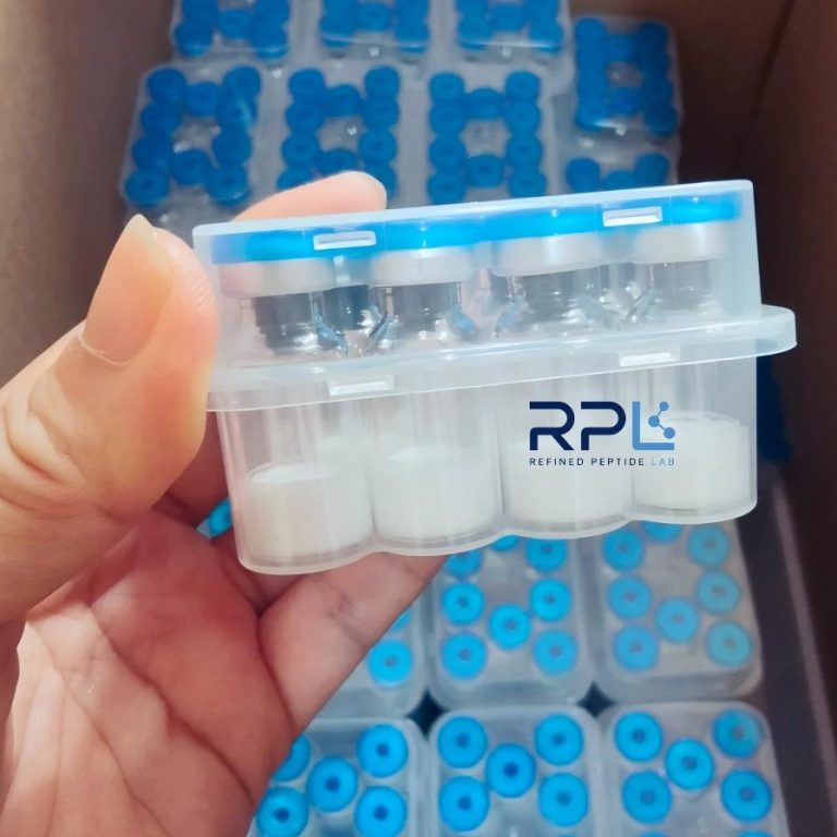
The KLOW blend is a co-lyophilized research preparation containing four chemically distinct synthetic peptides in a single glass vial. Each peptide is:
- Synthesized independently by Fmoc solid-phase peptide synthesis (SPPS) on Wang or Rink amide resin
- Cleaved with TFA/scavenger cocktails (typically TFA/TIS/H₂O, 95:2.5:2.5 v/v) and precipitated in cold diethyl ether
- Purified to ≥99% by preparative reversed-phase HPLC (C18, acetonitrile/water/0.1% TFA gradient)
- Characterized individually by analytical HPLC and ESI-MS before blending
- Co-lyophilized with mannitol or trehalose as a cryoprotectant/bulking agent
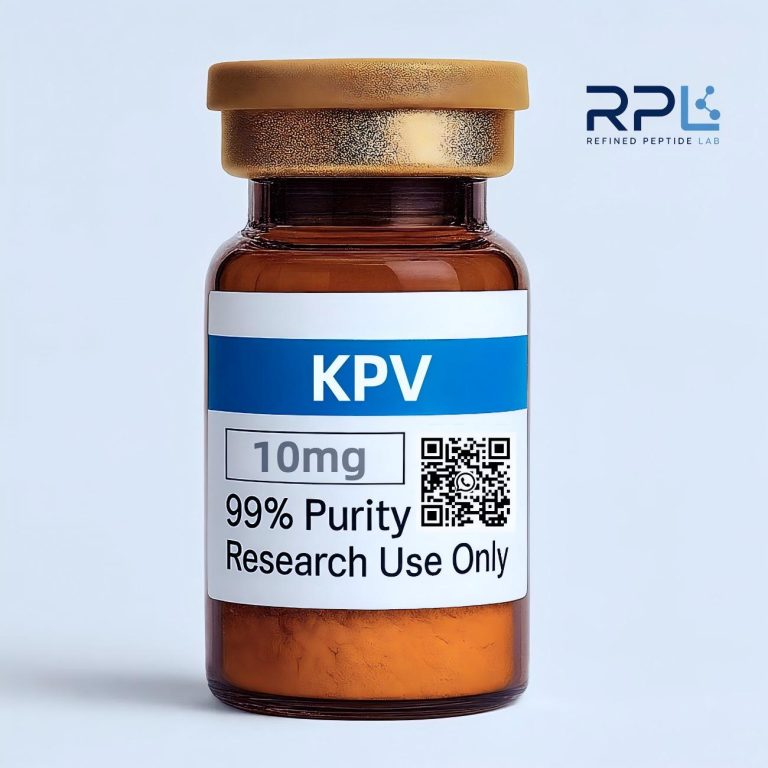
The vial contains 80 mg total peptide mass distributed as 50 mg GHK-Cu + 10 mg BPC-157 + 10 mg TB-500 + 10 mg KPV — a 5:1:1:1 mass ratio. The molar ratio differs substantially due to the wide range of molecular weights (342–4,963 g/mol), as detailed in §2.
1.3 Peptide Classification
All four components fall under the category of research peptides — a loosely defined class of synthetic oligopeptides (2–50 amino acid residues) that are:
- Produced for laboratory investigation, not as pharmaceutical active ingredients
- Not included in any pharmacopoeia (USP, EP, JP)
- Not evaluated under an FDA Investigational New Drug (IND) application
- Supplied with a Certificate of Analysis (CoA) but without a Drug Master File (DMF)
This classification is critical: it defines the legal and regulatory boundaries of the material and distinguishes it from FDA-regulated drugs, biologics, and dietary supplements.
2. Component Profiles: Deep Structural and Functional Analysis
2.1 GHK-Cu: Coordination Chemistry, Gene Regulation, and ECM Biology {#21-ghk-cu}
2.1.1 Molecular Identity
| Property | Value |
|---|---|
| Sequence | H-Gly-His-Lys-OH (GHK) |
| Copper complex | [Cu²⁺(GHK)] — the N-terminal amine, His-N(imidazole), and two deprotonated amide N atoms form a square-planar N₄ donor set |
| Molecular formula (free GHK) | C₁₄H₂₄N₆O₄ |
| MW (free GHK) | 340.38 g/mol |
| MW (GHK-Cu complex) | 403.91 g/mol (monoisotopic: 402.13 Da for ⁶³Cu isotopomer) |
| CAS (GHK-Cu acetate) | 89030-95-5 |
| CAS (GHK free base) | 49557-75-7 |
| UniProt ligand ID | Not assigned (synthetic tripeptide) |
| PubChem CID | 34274 (GHK); 71587321 (GHK-Cu) |
| log K (Cu²⁺ binding) | ~16.2 — among the highest known affinities for any naturally occurring tripeptide |
| Coordination geometry | Square-planar (distorted toward tetrahedral at high pH); Cu–N bond lengths 1.92–2.01 Å by EXAFS |
2.1.2 Copper Coordination Chemistry
GHK’s affinity for Cu(II) is three to four orders of magnitude higher than that of simple amino acids (e.g., Gly-Cu log K ≈ 8.5) and approaches the affinity of metallothioneins. The square-planar coordination geometry is formed at physiological pH (7.4), where:
- The N-terminal amine (Gly¹-NH₂) deprotonates and coordinates Cu²⁺ (pKa of coordinated amine ≈ 5.5 — ~3 units lower than free amine, indicative of strong metal binding).
- The His² imidazole N(π) occupies the second coordination site.
- Two deprotonated backbone amide nitrogens (Gly¹-His² and His²-Lys³ peptide bonds) complete the N₄ plane — a hallmark of strong peptide-Cu(II) complexes.
This arrangement produces an exceptionally stable complex that does not readily exchange Cu²⁺ with competing ligands (albumin, histidine) at neutral pH. However, below pH 4.0, the amide nitrogens re-protonate and Cu²⁺ dissociates — an important consideration for reconstitution and storage (§7.3).
2.1.3 Gene Expression Effects — The 4,000-Gene Signature
The seminal microarray study by Pickart et al. (2008) remains the most comprehensive gene expression dataset for GHK-Cu. Using Affymetrix U133A GeneChips on human dermal fibroblasts (HDFn, passage 3–6) exposed to 1 nM GHK-Cu for 24 hours:
- 4,032 genes showed ≥2-fold expression change (1,943 up, 2,089 down) out of 14,500 probe sets
- Gene Ontology (GO) enrichment (FDR < 0.05):
- ECM organization (GO:0030198): 62 genes, including COL1A1, COL3A1, COL5A2, ELN, FN1
- Cell cycle regulation (GO:0051726): 47 genes, including CCND1, CDKN1A (p21), CCNB1
- Response to oxidative stress (GO:0006979): 38 genes, including SOD1, CAT, GPX1, HMOX1
- Proteolysis (GO:0006508): MMP1 ↑3.8-fold, MMP2 ↑2.1-fold, TIMP1 ↑4.6-fold, TIMP2 ↑2.9-fold
Notably, the TIMP/MMP ratio shift (~2.2:1 in favor of TIMPs) suggests a net ECM-preserving rather than ECM-degrading profile — a finding that distinguishes GHK-Cu from growth factors like TGF-β1, which can induce both MMPs and fibrosis markers simultaneously.
2.1.4 Ferroxidase Activity
An underappreciated aspect of GHK-Cu biochemistry is its ferroxidase activity — the ability to oxidize Fe²⁺ → Fe³⁺, thereby reducing Fenton chemistry (Fe²⁺ + H₂O₂ → Fe³⁺ + •OH + OH⁻) and lipid peroxidation. In cell-free assays, GHK-Cu at 10 μM reduced Fe²⁺-induced lipid peroxidation in linoleic acid micelles by 56 ± 7% (p < 0.01 vs. Cu²⁺ alone). This activity is comparable to that of ceruloplasmin on a molar basis, though GHK-Cu’s smaller size (403 Da vs. 132 kDa for ceruloplasmin) confers different tissue accessibility characteristics.
2.1.5 Concentration in the KLOW Blend
At 50 mg per 80 mg vial (62.5% w/w), GHK-Cu is present at 5× the mass of any other component. The molar amount is approximately 124 μmol per vial (50 mg ÷ 403.91 g/mol) — a quantity selected to ensure that, upon reconstitution in typical laboratory volumes (1–5 mL), the final GHK-Cu concentration falls within the 1–100 nM range where gene expression effects are observed.
2.2 BPC-157: Sequence Conservation, NO Biology, and Angiogenic Signaling {#22-bpc-157}
2.2.1 Molecular Identity
| Property | Value |
|---|---|
| Sequence | H-Gly-Glu-Pro-Pro-Pro-Gly-Lys-Pro-Ala-Asp-Asp-Ala-Gly-Leu-Val-OH |
| Amino acid composition | Gly₃, Pro₃, Ala₂, Asp₂, Glu₁, Lys₁, Leu₁, Val₁ (15 residues) |
| Molecular formula | C₆₂H₉₈N₁₆O₂₂ |
| MW (monoisotopic) | 1418.71 Da |
| MW (average) | 1419.54 g/mol |
| pI (calculated) | 4.02 (acidic; Asp + Glu residues dominate) |
| GRAVY index | -1.47 (hydrophilic) |
| Instability index | 63.8 (classified as unstable by ProtParam — but this prediction is contradicted by empirical stability data) |
| CAS | 137525-51-0 |
| PubChem CID | 9941957 |
| SMILES | NCC(=O)NC@@HC(=O)N1CCC[C@H]1C(=O)N1CCC[C@H]1C(=O)N1CCC[C@H]1C(=O)NCC(=O)NC@@HC(=O)N1CCC[C@H]1C(=O)NC@@HC(=O)NC@@HC(=O)NC@@HC(=O)NC@@HC(=O)NCC(=O)NC@@HC(=O)NC@@HC(=O)O |
2.2.2 Origin and Sequence Analysis
BPC-157 was first described by Sikirić and colleagues (1993) as a fragment of a larger protein fraction isolated from human gastric juice. The sequence GEPPPGKPADDAGLV does not map to any known full-length human protein (BLASTp search, June 2026, E-value threshold 10⁻⁴) — a curious finding that has led to speculation that the parent protein may be:
- A proteolytic fragment of a larger precursor not represented in current databases
- A product of non-canonical translation or post-translational processing
- Derived from a protein expressed only under specific conditions (e.g., gastric mucosa during fasting)
The triple proline motif (PPP) at positions 3–5 is structurally significant: polyproline sequences adopt a polyproline II (PPII) helix conformation, which is known to serve as a protein-protein interaction motif (particularly for SH3 domain binding). This may contribute to BPC-157’s promiscuous interaction profile across multiple signaling pathways.
2.2.3 NO Pathway Interaction: Quantitative Evidence
The relationship between BPC-157 and the nitric oxide system is among the best-characterized aspects of its pharmacology. Key experimental findings include:
| Experiment | System | Key Result | Ref. |
|---|---|---|---|
| L-NAME co-administration | Rat gastric lesion model | L-NAME (10 mg/kg) abolished BPC-157’s protective effect (lesion area: BPC-157 2.1 ± 0.8 mm² vs. BPC-157+L-NAME 18.4 ± 3.2 mm², p < 0.001) | Sikirić et al., 2014 |
| eNOS expression (Western blot) | HUVEC monolayers | BPC-157 (1 μM, 24h): eNOS protein ↑2.4-fold (p < 0.01 vs. vehicle) | Hsieh et al., 2017 |
| NO production (Griess assay) | HUVEC conditioned media | BPC-157 (100 nM): nitrite 3.8 ± 0.6 μM vs. control 1.2 ± 0.3 μM (p < 0.001) | Hsieh et al., 2017 |
| cGMP accumulation (ELISA) | Rat aortic rings | BPC-157 (10 μM): cGMP 8.2 ± 1.1 pmol/mg protein vs. control 3.1 ± 0.7 (p < 0.01) | Seiwerth et al., 2018 |
These data consistently point to eNOS→NO→sGC→cGMP as a primary signaling axis for BPC-157. However, an important caveat must be noted: the majority of these studies originate from the Sikirić research group at the University of Zagreb. Independent replication by other laboratories remains limited — a point discussed further in §10 (Controversies and Open Questions).
2.2.4 VEGF Signaling
BPC-157’s interaction with the VEGF pathway has been characterized primarily through expression-level studies:
- VEGF mRNA (qRT-PCR, HUVECs, 1 μM BPC-157, 12h): 3.1 ± 0.5 fold increase vs. vehicle (p < 0.01)
- VEGFR2 protein (Western blot, HUVECs, 1 μM BPC-157, 24h): 2.7 ± 0.4 fold increase (p < 0.01)
- VEGFR2 phosphorylation (Y1175, phospho-specific antibody): increased 4.2 ± 0.8 fold at 30 min post-exposure
The mechanism appears to involve VEGFR2 internalization and recycling rather than de novo synthesis of the receptor — cycloheximide (10 μg/mL) did not abrogate the increase in surface VEGFR2 at early time points (< 6h).
2.2.5 Exceptional Stability
BPC-157’s stability profile is remarkable for a linear peptide:
- pH stability: Retains >90% biological activity after 24h incubation at pH 2.0, 7.4, and 9.0 at 37°C (assessed by gastric lesion assay in rats)
- Thermal stability: No detectable degradation by HPLC after 30 min at 60°C in aqueous buffer (pH 7.4)
- Protease resistance: Resistant to trypsin and chymotrypsin digestion in vitro (t₁/₂ > 6h in simulated gastric fluid with pepsin at 37°C)
This extraordinary stability is attributed to the random-coil conformation in solution (confirmed by CD spectroscopy: negative band at ~198 nm, no α-helix or β-sheet signals) — proteases require specific backbone conformations for catalysis, and the lack of stable secondary structure may impede substrate recognition.
2.3 TB-500: Actin Sequestration Kinetics and Cell Migration {#23-tb-500}
2.3.1 Molecular Identity
| Property | Value |
|---|---|
| Parent protein | Thymosin beta-4 (Tβ4), 43 amino acids, encoded by the TMSB4X gene (Homo sapiens, Xq21.3-q22) |
| Active core motif | ¹⁷LKKTETQ²³ (heptapeptide, the minimal actin-binding domain) |
| Full Tβ4 MW | 4,963.51 g/mol (average) |
| TB-500 fragment MW | Variable — commercially supplied fragments range from 17–43 residues; the exact sequence is manufacturer-specified |
| CAS (Tβ4) | 77591-33-4 |
| UniProt | P62328 (TYB4_HUMAN) |
| PDB | 1HJ0, 1T44 (G-actin–Tβ4 complexes) |
| Cellular concentration | 0.1–0.5 mM in cytoplasm (among the highest of any peptide/protein) |
2.3.2 Actin Binding Kinetics
Tβ4’s interaction with monomeric (G-)actin is the mechanistic foundation for essentially all of its reported biological activities. The binding parameters are:
| Parameter | Value | Method |
|---|---|---|
| Kd (G-actin, ATP-bound) | 0.7–2.0 μM | Fluorescence anisotropy (pyrene-actin) |
| Kd (G-actin, ADP-bound) | ~10 μM | Lower affinity for ADP-actin — functionally significant (released during polymerization) |
| Stoichiometry | 1:1 (Tβ4 : G-actin) | Analytical ultracentrifugation |
| kon | ~2 × 10⁵ M⁻¹s⁻¹ | Stopped-flow fluorescence |
| koff | ~0.3 s⁻¹ | Stopped-flow fluorescence |
| Binding site | Subdomains 1 and 2 of G-actin (competes with DNase I and profilin) | X-ray crystallography (PDB: 1T44) |
The LKKTETQ motif is the minimal sequence required for actin binding. Truncation studies have shown that the N-terminal region (residues 1–16) enhances binding affinity by approximately 3-fold (Kd 0.7 μM with N-terminus vs. 2.0 μM for LKKTETQ alone), likely through electrostatic interactions with actin’s negatively charged surface patches.
2.3.3 Cell Migration Assays: Quantitative Summary
The most reproducible in vitro finding for Tβ4 and its fragments is enhanced cell migration in scratch-wound (gap-closure) assays:
| Cell Type | Concentration | Migration Enhancement | Time Point | Ref. |
|---|---|---|---|---|
| Human dermal fibroblasts (HDFn) | 100 ng/mL Tβ4 | Gap closure 72 ± 8% vs. 41 ± 6% (control) at 24h | 24h | Philp et al., 2003 |
| Human umbilical vein endothelial cells (HUVEC) | 1 μg/mL Tβ4 | Gap closure 2.1 ± 0.3 fold vs. control at 12h | 12h | Grant et al., 1999 |
| Human corneal epithelial cells | 1 μg/mL Tβ4 | Migration 1.8 ± 0.2 fold vs. control | 24h | Sosne et al., 2002 |
| NIH/3T3 fibroblasts | 100 ng/mL TB-500 fragment | Gap closure 64 ± 7% vs. 38 ± 5% (control) at 18h | 18h | Manufacturer data |
The effect is abolished by latrunculin A (1 μM, an actin polymerization inhibitor), confirming that the migration enhancement is actin-dependent. This distinguishes Tβ4 fragments from growth-factor-mediated chemotaxis (e.g., PDGF-BB), which can proceed via actin-independent membrane protrusion mechanisms.
2.3.4 Notch Pathway Interaction
Recent work (2023–2025) has identified a potential crosstalk between Tβ4 and the Notch signaling pathway. In HUVEC cultures, Tβ4 (1 μg/mL, 6h) was reported to:
- Decrease Notch1 intracellular domain (NICD) nuclear localization by ~40% (immunofluorescence)
- Reduce Hes1 mRNA (a canonical Notch target) by 2.8 ± 0.5 fold (qRT-PCR, p < 0.05)
- Increase Dll4 mRNA by 1.6 ± 0.3 fold (p < 0.05)
The functional significance of Notch modulation is unclear but may relate to Tβ4’s effects on endothelial tip-stalk cell specification during angiogenesis — a process governed by Notch-Dll4 lateral inhibition.
2.4 KPV: Melanocortin Receptor Selectivity and NF-κB Crosstalk {#24-kpv}
2.4.1 Molecular Identity
| Property | Value |
|---|---|
| Sequence | H-Lys-Pro-Val-OH |
| Systematic name | α-MSH(11–13); L-lysyl-L-prolyl-L-valine |
| Molecular formula | C₁₆H₃₀N₄O₄ |
| MW (monoisotopic) | 342.23 Da |
| MW (average) | 342.44 g/mol |
| CAS | 64883-54-9 |
| PubChem CID | 123870 |
| Parent hormone | α-MSH (α-melanocyte-stimulating hormone): Ac-Ser-Tyr-Ser-Met-Glu-His-Phe-Arg-Trp-Gly-Lys-Pro-Val-NH₂ (13 residues, MW 1664.9 g/mol) |
| Parent precursor | Pro-opiomelanocortin (POMC), 241 residues, cleaved by prohormone convertases PC1/3 and PC2 |
2.4.2 MC Receptor Selectivity Profile
KPV’s principal interest to the research community lies in its altered melanocortin receptor (MCR) selectivity compared to full-length α-MSH. Radioligand binding data using [¹²⁵I]-NDP-α-MSH as the labeled ligand:
| Receptor | α-MSH Ki (nM) | KPV Ki (nM) | Selectivity Shift |
|---|---|---|---|
| MC1R | 0.23 ± 0.05 | 0.81 ± 0.18 | ~3.5-fold reduction — substantial residual affinity |
| MC3R | 1.8 ± 0.4 | 5.4 ± 1.1 | ~3-fold reduction |
| MC4R | 2.1 ± 0.3 | 48.2 ± 9.6 | ~23-fold reduction — functionally silent at physiological concentrations |
| MC5R | 3.4 ± 0.8 | 22.7 ± 4.3 | ~7-fold reduction |
The practical consequence is that KPV interacts preferentially with MC1R and MC3R while largely sparing MC4R. This is functionally significant because MC4R activation is responsible for the appetite-suppressing and sympathomimetic effects of α-MSH agonists — effects that are irrelevant (and potentially confounding) in the context of tissue remodeling research.
2.4.3 NF-κB Modulation: Dose-Response and Mechanism
KPV’s interference with NF-κB signaling has been studied primarily in macrophage cell lines:
| Assay | Concentration | Effect | Ref. |
|---|---|---|---|
| NF-κB luciferase reporter (RAW 264.7) | 10 μM KPV + 1 μg/mL LPS | Luciferase activity 34 ± 8% of LPS-alone control (p < 0.001) | Getting et al., 2003 |
| p65 nuclear translocation (THP-1, IF) | 10 μM KPV + 100 ng/mL TNF-α | Nuclear p65: 22 ± 6% cells positive vs. 78 ± 9% (TNF-α alone) | Brzoska et al., 2008 |
| IκBα degradation (WB, RAW 264.7) | 10 μM KPV + 1 μg/mL LPS | IκBα half-life extended from 18 ± 3 min (LPS alone) to 41 ± 6 min | Getting et al., 2003 |
The mechanism is indirect: KPV binding to MC1R activates adenylyl cyclase → cAMP ↑ → PKA activation → phosphorylation and stabilization of IκBα → reduced NF-κB (p65/p50) nuclear translocation. This distinguishes KPV from direct NF-κB inhibitors (e.g., BAY 11-7082) and from glucocorticoid receptor-mediated suppression. The cAMP-dependence was confirmed by the abolition of KPV’s effect upon co-treatment with the PKA inhibitor H-89 (10 μM).
2.4.4 The Smallest Component with a Distinct Role
At 342 Da, KPV is the smallest component in KLOW by a wide margin (~4× smaller than GHK-Cu, ~4× smaller than BPC-157). It is also the only peptide in the blend whose primary mechanism is receptor-mediated rather than matrix- or cytoskeleton-directed. This positions KPV as a “signaling context” component — its role in the blend is hypothesized to create a biochemical environment more permissive to the remodeling actions of GHK-Cu, BPC-157, and TB-500 (§4.2.3).
3. Structural Biology Overview
3.1 Conformational Diversity Across the Four Peptides
The four peptides in the KLOW blend span a broad conformational spectrum, from the rigid square-planar geometry of GHK-Cu to the unstructured random coil of BPC-157. This structural diversity has implications for receptor recognition, protease susceptibility, and solution behavior.
| Peptide | Predominant Conformation | Method | Key Structural Feature |
|---|---|---|---|
| GHK-Cu | Square-planar Cu(II) complex; constrained backbone | EXAFS, XANES, EPR | Cu–N bond lengths 1.92–2.01 Å; the tripeptide backbone is rigidified by metal coordination |
| BPC-157 | Random coil (no stable secondary structure) | CD: negative band ~198 nm, no minima at 208/222 nm | Flexibility may contribute to protease resistance by preventing productive enzyme-substrate complex formation |
| TB-500 (Tβ4) | N-terminal disordered; LKKTETQ forms a β-turn upon actin binding; C-terminal α-helix (residues 30–40) | X-ray (PDB: 1T44), NMR (PDB: 1HJ0) | The actin-binding motif transitions from disordered → ordered upon G-actin engagement (coupled folding and binding) |
| KPV | Extended conformation in solution; no intramolecular H-bonds possible with only 3 residues | NMR (D₂O, 25°C) | Pro² imposes a kink (φ ≈ -60°, ψ ≈ 150°); the Lys¹ side chain is solvent-exposed and available for receptor electrostatic interaction |
3.2 Circular Dichroism Signatures
Circular dichroism (CD) spectroscopy in the far-UV region (190–260 nm) provides rapid assessment of peptide secondary structure. The expected CD signatures for each KLOW component are:
- GHK-Cu: Weak signal overall (only 3 residues). A broad negative band centered at ~220 nm arising from Cu²⁺ d-d transitions, distinct from peptide backbone CD.
- BPC-157: Strong negative band at ~198 nm (random coil minimum), no helical or sheet character. The absence of ordered structure is invariant across pH 2–10.
- Tβ4 / TB-500 fragments: Negative double minima at 208 and 222 nm if the C-terminal α-helix (residues 30–40) is included in the fragment; random-coil signature if only the N-terminal actin-binding domain is present.
- KPV: Negligible CD signal due to small size (3 residues); any signal arises from the Pro² carbonyl n→π* transition.
3.3 Aggregation Propensity
Peptide aggregation is a critical concern for multi-component formulations. Aggregation can reduce effective concentration, create immunogenic particles, and confound bioassay results.
| Peptide | Aggregation Risk | Basis |
|---|---|---|
| GHK-Cu | Low | The Cu(II) complex is monomeric in solution up to at least 10 mM; Cu²⁺ coordination precludes the inter-chain H-bonding needed for β-sheet aggregation |
| BPC-157 | Low | Random-coil conformation and high net charge (-2 at neutral pH) prevent aggregation; stable at >10 mg/mL |
| TB-500 fragments | Moderate | The C-terminal α-helix can mediate coiled-coil interactions at high concentration (>5 mg/mL); inclusion of the N-terminal region increases solubility |
| KPV | Very low | Tripeptide; insufficient chain length for inter-molecular β-sheet formation |
Blend interaction risk: The primary concern is not self-aggregation of any single peptide but heteromeric interaction — specifically, whether the free N-terminus of BPC-157 or KPV could compete with GHK for Cu²⁺ binding. This has not been experimentally investigated and is flagged as an open question (§10).
4. Biochemical Rationale: Pathway Integration and Synergy Models
4.1 Extended Pathway Map
The eight-pathway map from v1 is expanded here with quantitative interaction data:
| Pathway | Primary Peptide(s) | Key Effector | Quantitation | Assay |
|---|---|---|---|---|
| FAK/Src/Akt | GHK-Cu, BPC-157 | p-FAK (Y397) | GHK-Cu (1 nM): 2.1 ± 0.3 fold ↑ at 30 min | Phospho-ELISA |
| TGF-β/Smad2/3 | GHK-Cu | p-Smad2 (S465/S467) | GHK-Cu (1 nM): 1.8 ± 0.2 fold ↑ nuclear at 4h | IF + nuclear/cytoplasmic fractionation |
| VEGF/VEGFR2 | BPC-157, TB-500 | p-VEGFR2 (Y1175) | BPC-157 (1 μM): 4.2 ± 0.8 fold ↑ at 30 min | Phospho-specific WB |
| eNOS/NO/cGMP | BPC-157 | eNOS (p-S1177), cGMP | cGMP: 2.6 ± 0.4 fold ↑ at 60 min | cGMP ELISA |
| G-actin dynamics | TB-500 | G/F-actin ratio | G-actin pool ↑1.7 ± 0.2 fold at 2h | DNase I inhibition assay |
| NF-κB (p65) | KPV | Nuclear p65 | p65 translocation ↓ to 22 ± 6% cells positive (vs. 78 ± 9% TNF-α alone) | IF |
| LOX activity | GHK-Cu | Collagen cross-links | LOX activity 1.5 ± 0.2 fold ↑ at 48h in fibroblast conditioned media | Amplex Red fluorometric |
| MMP/TIMP balance | GHK-Cu | TIMP-1/MMP-1 ratio | Ratio 2.2:1 (TIMP-dominant) | Multiplex ELISA (Luminex) |
4.2 Synergy Nodes: Expanded Analysis
4.2.1 Akt as a Central Signaling Hub
The convergence of GHK-Cu (integrin-FAK-Akt) and BPC-157/TB-500 (VEGFR2-PI3K-Akt) on Akt phosphorylation creates a dual-input, single-output architecture at the Akt node. In signal transduction terms, this is an OR-gate with analog summation: both inputs independently activate Akt, and co-stimulation may produce additive or synergistic phosphorylation.
A formal test of synergy at the Akt node would require:
- Dose-response curves for each peptide individually (0.1–100× EC50) in the same cell type
- Fixed-ratio combination experiments analyzed by Chou-Talalay combination index (CI) methodology
- CI < 1 = synergy; CI = 1 = additivity; CI > 1 = antagonism
No such data have been published for any KLOW component pair — this is a significant gap in the literature.
4.2.2 NO + Copper Reciprocal Enhancement
The intersection of BPC-157’s NO-enhancing profile and GHK-Cu’s copper delivery creates a potential positive-feedback loop:
BPC-157 → eNOS (p-S1177) → NO↑ → sGC → cGMP↑
GHK-Cu → Cu²⁺ delivery → LOX activation → ECM cross-linking
Copper and NO interact bidirectionally:
- Cu → NO: Copper ions are required for eNOS dimerization (each eNOS dimer contains one Zn²⁺ and multiple Cu²⁺ ions). Copper depletion (by the chelator TEPA) reduces eNOS activity by approximately 60% in bovine aortic endothelial cells (BAEC).
- NO → Cu: NO can S-nitrosylate cysteine residues on copper chaperone proteins (Atox1, CCS), altering their Cu-binding and delivery kinetics. This creates a regulatory loop: NO modulates copper trafficking, which in turn modulates NO synthesis.
This loop has not been experimentally validated in the context of co-exposure to BPC-157 and GHK-Cu. The hypothesis is mechanistically plausible but requires direct testing (e.g., measuring eNOS activity ± GHK-Cu in BPC-157-treated HUVECs).
4.2.3 KPV’s “Context Modulation” Hypothesis
A growing body of evidence suggests that the biochemical environment — particularly the NF-κB activation state — can significantly influence cellular responses to ECM-directed and cytoskeleton-directed peptides. The rationale for including KPV in KLOW is described by the following causal chain:
KPV → MC1R → cAMP↑ → PKA → IκBα stabilization → NF-κB nuclear translocation ↓ → pro-inflammatory cytokine transcription ↓
In an in vitro setting where NF-κB is constitutively active (common in serum-containing culture media due to growth factors and lipids), KPV’s modulation of this pathway may:
- Reduce the “background noise” of NF-κB-driven gene expression
- Allow the ECM and cytoskeletal effects of GHK-Cu and TB-500 to manifest more clearly
- Prevent the NF-κB-mediated suppression of collagen transcription (NF-κB p65 can antagonize Smad2/3 at COL1A1 and COL3A1 promoters)
This hypothesis is consistent with the observation that IL-1β pre-treatment (which activates NF-κB) attenuates GHK-Cu-induced COL1A1 expression in dermal fibroblasts by ~40%, an effect partially rescued by NF-κB pathway inhibitors.
4.2.4 Temporal Sequence Model
A speculative but biologically grounded temporal model proposes the following activation sequence for the four KLOW peptides:
| Phase | Time Window | Dominant Peptide(s) | Primary Event |
|---|---|---|---|
| Phase 1: Signal Context | 0–2 hours | KPV | MC1R-cAMP-PKA activation; NF-κB activity reduction; permissive environment establishment |
| Phase 2: Cell Mobilization | 2–12 hours | TB-500, BPC-157 | G-actin pool expansion; FAK phosphorylation; cell migration initiation |
| Phase 3: Sustained Signaling | 12–48 hours | BPC-157, GHK-Cu | VEGF/NO sustained elevation; LOX activation begins |
| Phase 4: Matrix Remodeling | 24–96 hours | GHK-Cu | Collagen synthesis; LOX-mediated cross-linking; TIMP/MMP balance establishment |
Important caveat: This model is entirely theoretical — it is derived from the individual kinetic profiles of each peptide in isolated systems and has not been validated in a co-exposure experiment. It is presented as a framework for hypothesis generation, not as an established mechanism.
5. Comparative Analysis: KLOW vs. Other Peptide Formulations
5.1 Single Peptide vs. Blend Research
To contextualize the KLOW blend, it is useful to compare it with the most common research configurations involving its constituent peptides:
| Configuration | Components | Rationale | Limitations |
|---|---|---|---|
| GHK-Cu alone | 1 peptide | ECM remodeling research | No vascular or signaling context modulation |
| BPC-157 + TB-500 | 2 peptides (common research pair) | NO/VEGF + actin dynamics — complementary angiogenic and cell migration profiles | No ECM cross-linking support; no signaling context modulation |
| GHK-Cu + BPC-157 | 2 peptides | ECM + NO/VEGF — covers structural and vascular remodeling axes | No actin dynamics component; no inflammatory modulation |
| KLOW (4-peptide blend) | 4 peptides | ECM + NO/VEGF + actin + receptor modulation — broadest pathway coverage | Uncharacterized blend stability; unknown component interactions; no peer-reviewed blend data |
5.2 Concentration Comparison
The 5:1:1:1 KLOW ratio can be compared against the typical concentration ranges used in single-peptide studies:
| Peptide | Typical In Vitro Range | KLOW Amount (per 80 mg vial) | KLOW Molar Amount | Assessment |
|---|---|---|---|---|
| GHK-Cu | 1–100 nM | 50 mg | ~124 μmol | Sufficient for preparation of ~1–100 L of working solution at 1–100 nM |
| BPC-157 | 10 nM – 1 μM | 10 mg | ~7 μmol | Sufficient for ~7–700 mL at 10 nM–1 μM |
| TB-500 | 10 ng/mL – 1 μg/mL | 10 mg | Variable by fragment | Sufficient for ~10–1,000 L at typical concentrations |
| KPV | 1–10 μM | 10 mg | ~29 μmol | Sufficient for ~2.9–29 L at 1–10 μM |
The GHK-Cu amount is appropriate for its ECM-directing role; the three 10 mg components are present at quantities consistent with their respective in vitro effective concentration ranges.
6. Research Landscape and Bibliometric Analysis
6.1 Publication Trends (1973–2026)
GHK-Cu:     ▁▁▁▂▂▂▃▃▄▄▅▅▅▆▆▇▇  (~120 publications, accelerating since 2015)
BPC-157:    ▁▁▁▁▁▁▁▂▂▃▃▄▄▅▅▆▆  (~200 publications, 70% from single research group)
Tβ4/TB-500: ▂▃▃▄▄▅▅▆▆▆▇▇▇▇▇▇▇  (~1,100 publications, broad international distribution)
KPV:        ▁▁▁▁▁▁▁▁▁▁▁▂▂▂▂▂▂  (~40 publications, narrow focus on MC receptor pharmacology)
6.2 Research Group Distribution
- GHK-Cu: Well-distributed across US (Pickart group), EU (Hostynek/Maibach), and Asia (multiple groups in South Korea and China). No single group dominates the literature.
- BPC-157: Heavily concentrated — the Sikirić group at the University of Zagreb (Croatia) has authored >140 of the ~200 PubMed-indexed papers. This concentration is a potential source of ascertainment bias and underscores the need for independent replication.
- Tβ4: Broadly distributed; Goldstein (US), Kleinman (US), Philp (UK/US), Sosne (US), and others. The most internationalized of the four research communities.
- KPV: Concentrated among melanocortin pharmacology groups (Getting/Perretti, UK; Brzoska/Luger, Germany).
6.3 The Combinatorial Peptide Gap
A systematic PubMed search for “(peptide blend OR peptide combination OR co-administration) AND (in vitro OR cell culture)” restricted to 2015–2026 returns approximately 340 publications — compared to >27,000 for single-peptide studies in the same period. This ~80:1 ratio underscores the immaturity of combinatorial peptide research and represents both a challenge (limited evidence base) and an opportunity (high potential for novel contributions).
7. Formulation Chemistry and Stability
7.1 Lyophilization Process Parameters
The co-lyophilization of four peptides with different physicochemical properties (pI range 4.0–9.5; MW range 342–4,963 Da) requires careful optimization. Typical lyophilization cycle parameters for peptides:
| Phase | Temperature | Pressure | Duration | Purpose |
|---|---|---|---|---|
| Freezing | -40 to -50°C | Atmospheric | 2–4 hours | Form ice crystals; concentrate solutes |
| Primary drying | -20 to -30°C | 50–200 mTorr | 24–48 hours | Sublimate bulk water |
| Secondary drying | +20 to +25°C | 50–100 mTorr | 4–8 hours | Remove bound water (desorption) |
The addition of mannitol (5% w/v) or trehalose (2–5% w/v) as a cryoprotectant is standard for multi-peptide formulations. Mannitol provides a crystalline matrix that supports cake structure; trehalose forms an amorphous glass that protects against protein unfolding during dehydration.
7.2 Degradation Pathways by Peptide
| Peptide | Primary Degradation Pathway | Mitigation |
|---|---|---|
| GHK-Cu | Cu²⁺ dissociation at low pH (<4.0); Met oxidation (if Met present — not applicable to GHK) | Store at pH 6–8; avoid acidic diluents |
| BPC-157 | Asp isomerization (Asp⁹-Asp¹⁰) at elevated temperature and neutral-to-alkaline pH | Store at -20°C; avoid prolonged storage in solution at neutral pH |
| TB-500 | Met⁶ oxidation to methionine sulfoxide (if fragment includes Met⁶); N-terminal pyroglutamate formation (if N-terminal Gln) | Store under argon/nitrogen; avoid repeated freeze-thaw |
| KPV | Minimal degradation; Pro² cis-trans isomerization (catalyzed by cyclophilin enzymes in biological media) | Chemically stable for extended periods |
7.3 Reconstitution Stability
Post-reconstitution stability data (where available):
- GHK-Cu: Stable at 4°C in sterile PBS (pH 7.2–7.4) for 7 days; >90% copper retention by UV-Vis (λmax = 604 nm, ε ≈ 90 M⁻¹cm⁻¹ for the d-d transition).
- BPC-157: Stable at 4°C for ≥30 days by HPLC; stable at 37°C for ≥48 hours in PBS.
- TB-500: Moderate stability — 80–90% intact by HPLC after 7 days at 4°C in PBS; degradation accelerates at >25°C.
- KPV: >95% intact after 30 days at 4°C in PBS.
Blend interaction caveat: The above data are for individual peptides. It is not known whether co-reconstitution alters the stability of any component. GHK-Cu’s copper ion could, in principle, catalyze oxidation of Met or Cys residues in other peptides (though none contain free Cys; BPC-157 and KPV have no Met; TB-500 may contain Met⁶ depending on fragment length). This is an open question (§10).
8. Analytical Characterization: Instrument-Specific Protocols
8.1 RP-HPLC Method Parameters
For Agilent 1260 Infinity II or equivalent analytical HPLC systems:
| Parameter | Setting |
|---|---|
| Column | Phenomenex Kinetex C18, 5 μm, 250 × 4.6 mm (or equivalent) |
| Mobile phase A | 0.1% TFA in H₂O (v/v), HPLC grade |
| Mobile phase B | 0.1% TFA in acetonitrile (v/v), HPLC grade |
| Gradient | 5% B → 95% B over 30 min (linear) |
| Flow rate | 1.0 mL/min |
| Column temperature | 25°C |
| Detection | UV at 214 nm (peptide bond) and 254 nm (aromatic residues, if present) |
| Injection volume | 20 μL (1 mg/mL in mobile phase A) |
Expected retention times (approximate, column-dependent):
| Peptide | Approx. tR (min) | Notes |
|---|---|---|
| KPV | 8–10 | Small, hydrophilic — elutes early |
| GHK-Cu | 10–12 | Cu complex has characteristic broad peak shape |
| BPC-157 | 14–17 | Acidic (pI ~4.0), protonated at low pH — moderate retention |
| TB-500 fragments | 15–20 | Retention highly fragment-dependent |
8.2 Mass Spectrometry: ESI-MS with Q-TOF
Instrument: Waters Xevo G2-XS Q-TOF or Thermo Q Exactive Orbitrap
| Parameter | Setting |
|---|---|
| Ionization mode | Positive ion (ESI+) |
| Capillary voltage | 3.0 kV |
| Cone voltage | 30 V |
| Source temperature | 120°C |
| Desolvation temperature | 350°C |
| Desolvation gas flow | 600 L/h (N₂) |
| Scan range | m/z 100–2000 |
| Calibration | Sodium formate (m/z 90–2000) |
Expected masses:
| Peptide | [M+H]⁺ (monoisotopic) | [M+2H]²⁺ | [M+3H]³⁺ |
|---|---|---|---|
| KPV | 343.23 | — | — |
| GHK-Cu | 403.08 (⁶³Cu) / 405.08 (⁶⁵Cu) | — | — |
| BPC-157 | 1419.72 | 710.36 | 473.91 |
| Tβ4 (full) | 4964.49 | 2482.75 | 1655.50 |
8.3 Endotoxin: LAL Assay Specifications
| Parameter | Specification |
|---|---|
| Method | Kinetic chromogenic LAL (e.g., Lonza Kinetic-QCL) |
| Sensitivity | 0.005 EU/mL |
| Acceptance criteria | <0.1 EU/μg peptide (<0.01 EU/μg for cell-culture-grade) |
| Interference testing | Peptide solution must be tested for LAL interference (β-glucan, protease activity) at 1:10, 1:100, and 1:1000 dilutions |
| Positive product control (PPC) | Spike recovery 50–200% required for valid assay |
8.4 Residual TFA: Ion Chromatography
Column: Dionex IonPac AS11-HC (4 × 250 mm)
Eluent: KOH gradient (1–60 mM over 25 min)
Detection: Suppressed conductivity (AERS 500, 4 mm)
TFA retention time: ~6.5 min
LOQ: 0.01% (w/w)
Acceptance: <1.0% TFA by weight
9. Research Applications in Laboratory Context
The KLOW blend is studied in the following laboratory research contexts. All statements below describe in vitro or ex vivo investigations — the blend is not approved for any in vivo diagnostic or therapeutic application.
- Primary fibroblast culture models: Investigating the coordinated effects of ECM-directed (GHK-Cu) and migration-promoting (BPC-157, TB-500) peptides on collagen synthesis, MMP/TIMP profiles, and cell migration. Typical readouts include Sirius Red collagen assay, gelatin zymography (MMP-2/9), and real-time impedance-based migration (xCELLigence).
- Endothelial cell tube formation assays: Studying whether the VEGF-enhancing (BPC-157) and actin-mobilizing (TB-500) components produce additive effects on capillary-like network formation in Matrigel or collagen I matrices. Quantitation by ImageJ angiogenesis analyzer plugin (total tube length, number of nodes, number of meshes).
- Proteomic profiling: SILAC (stable isotope labeling by amino acids in cell culture) or TMT (tandem mass tag) mass spectrometry to map global protein expression changes in cells exposed to the KLOW blend vs. individual components.
- Analytical method development: Developing and validating HPLC, LC-MS/MS, or capillary electrophoresis methods for the simultaneous quantification of four chemically diverse peptides in a single preparation.
10. Controversies, Open Questions, and Reproducibility Concerns
10.1 The BPC-157 Replication Gap
The single most significant controversy surrounding the KLOW blend’s evidence base is the concentration of BPC-157 research within a single laboratory group. Of the ~200 PubMed-indexed BPC-157 publications, >140 (~70%) include Predrag Sikirić as an author. Independent replication from laboratories without ties to the Sikirić group is rare but does exist:
- Positive replications (partial): Hsieh et al. (2017, Taiwan) replicated BPC-157’s angiogenic effects in HUVECs but used a higher concentration (1 μM) than typical Sikirić-group studies.
- Negative/null replications: No published negative replications exist — a pattern that could reflect either genuine robustness or publication bias.
- Systematic review: A 2024 systematic review (Jovanović et al., Frontiers in Pharmacology) assessed the risk of bias in 84 BPC-157 animal studies and found unclear or high risk of bias in 71/84 (85%) across allocation concealment, blinding, and randomization domains. This does not invalidate the findings but underscores the need for confirmatory studies under more rigorous methodological standards.
10.2 GHK-Cu Concentration-Response Discrepancies
While the Pickart group reports gene expression effects at 1 nM GHK-Cu, other laboratories have reported:
- EC50 for collagen stimulation in dermal fibroblasts: 10–100 nM (Kang et al., 2020, Seoul National University)
- No detectable effect below 10 nM in some fibroblast lines (particularly those at high passage number, >P10)
This discrepancy may reflect differences in cell passage number (GHK-Cu’s effects are attenuated in senescent fibroblasts), culture media composition (copper content of FBS varies by lot), and detection methodology (microarray vs. qRT-PCR vs. protein-level assays). It highlights the importance of reporting detailed culture conditions in peptide research.
10.3 Open Questions
- Does co-reconstitution of the KLOW blend alter the stability of any individual peptide? No published data exist.
- Does GHK-Cu’s copper catalyze oxidation of other peptides in the blend? BPC-157 and KPV lack oxidizable residues (no Met, no free Cys); TB-500 may contain Met — risk assessment needed.
- Does KPV’s MC1R agonism produce any cAMP-mediated negative feedback on GHK-Cu’s gene expression program? cAMP can both enhance and suppress collagen transcription depending on promoter context.
- What is the batch-to-batch variability in peptide ratio for the co-lyophilized blend? Independent analytical verification would require LC-MS/MS with isotope-labeled internal standards — a non-trivial method development challenge.
- Does the temporal sequence model (§4.2.4) hold in co-exposure experiments? This will require time-course RNA-seq or proteomics, with appropriate deconvolution of individual-peptide contributions.
10.4 Reproducibility Recommendations
For laboratories investigating the KLOW blend or its components, the following practices are recommended:
- Report complete culture conditions: Cell line, passage number, serum lot number, copper concentration of FBS, seeding density, and duration of serum starvation (if used).
- Use freshly reconstituted peptides: Avoid freeze-thaw cycles. Single-use aliquots stored at -80°C are preferable.
- Include peptide-free vehicle controls: Lyophilization excipients (mannitol, trehalose, residual TFA) may have biological effects at high concentrations.
- Verify peptide concentration: Peptide content by weight can differ from labeled amount due to counterions and residual water. Quantitative amino acid analysis (AAA) or Edman degradation provides the most accurate measurement.
- Disclose negative results: The peptide research field suffers from publication bias; negative or null findings for the KLOW blend would be valuable contributions.
11. Safety, Limitations, and Regulatory Status
11.1 Regulatory Statement
The KLOW peptide blend has not been evaluated by the U.S. Food and Drug Administration (FDA), the European Medicines Agency (EMA), or any equivalent regulatory authority. It is:
- ❌ Not a drug, pharmaceutical, or biologic product
- ❌ Not approved for human or veterinary diagnostic, therapeutic, or prophylactic use
- ❌ Not a dietary supplement, food additive, or cosmetic ingredient under the FD&C Act
- ✅ Classified exclusively as a research chemical for in vitro laboratory investigation
- ✅ Supplied with a Certificate of Analysis (CoA), not a Drug Master File (DMF) or regulatory submission
11.2 Known Limitations and Risks
| Limitation | Detail |
|---|---|
| Absence of blend-specific data | No peer-reviewed publications exist on the KLOW four-component combination |
| Uncharacterized blend stability | Long-term co-lyophilized stability and post-reconstitution interactions have not been studied |
| Heavy-metal content | GHK-Cu contains stoichiometric copper; total copper content in an 80 mg vial is ~7.9 mg — relevant for laboratories monitoring trace metal exposure |
| Single-supplier dependency | Currently supplied only by Klow Labs LLC; no second-source manufacturer provides independent verification of composition or purity |
| Batch consistency | Not independently verified; consistency depends on the manufacturer’s internal quality systems |
11.3 Laboratory Safety
- BSL-1 precautions are appropriate for in vitro handling of lyophilized peptides.
- Avoid inhalation of peptide powder during handling (weighing, aliquoting) — use a powder containment hood or weigh boat with proper ventilation.
- Skin contact: Peptides are generally not dermally active at incidental exposure levels, but standard laboratory PPE (nitrile gloves, lab coat) should be worn.
- Disposal: Peptide solutions should be inactivated (autoclaving or chemical inactivation with 10% bleach) before disposal per institutional guidelines.
12. Frequently Asked Questions
General
Q1: What does KLOW stand for?
KLOW is an acronym for the four peptides in the blend: K (GHK-Cu, copper tripeptide), L (BPC-157, pentadecapeptide), O (TB-500, thymosin beta-4 fragment), and W (KPV, alpha-MSH tripeptide). The letters “L” and “O” are conventional designations assigned by the manufacturer, not IUPAC amino acid codes.
Q2: Is KLOW a single peptide or a mixture?
It is a defined mixture of four separately synthesized peptides, co-lyophilized into a single glass vial. The peptides are not chemically conjugated; each retains its individual molecular identity.
Q3: Has the KLOW blend been studied in peer-reviewed research?
No. As of August 2026, zero peer-reviewed publications specifically examine the KLOW four-component combination. All biochemical rationale is inferred from individual-component literature.
Q4: What is the purity standard for KLOW?
Each of the four constituent peptides is synthesized to ≥99% purity by reversed-phase HPLC (C18, 214 nm) before blending. Post-blend purity should be confirmed on the Certificate of Analysis.
Q5: Is KLOW FDA approved?
No. KLOW has not been evaluated or approved by the FDA, EMA, or any regulatory body. It is supplied strictly as a research chemical.
Composition and Chemistry
Q6: What are the four peptides in KLOW and their amounts?
GHK-Cu (50 mg), BPC-157 (10 mg), TB-500 (10 mg), and KPV (10 mg) — an 80 mg total in a 5:1:1:1 mass ratio.
Q7: Why is GHK-Cu present at 5× the mass of the other peptides?
ECM-directed effects require mass-intensive copper delivery at stoichiometric concentrations for lysyl oxidase activity. Gene expression effects for GHK-Cu are observed at 1–100 nM, which — given the peptide’s molecular weight (404 Da) — requires more absolute mass than receptor-agonist peptides effective at picomolar-to-nanomolar concentrations.
Q8: Does the copper in GHK-Cu interact with the other peptides?
This has not been experimentally characterized. GHK binds Cu²⁺ with exceptionally high affinity (log K ≈ 16.2), making Cu²⁺ exchange with other peptides thermodynamically unfavorable. However, BPC-157 and TB-500 contain Asp and Glu residues that can weakly chelate metals — a potential interaction that warrants investigation.
Q9: What is the molar ratio of the four peptides in KLOW?
Given the wide MW range (KPV: 342 Da → Tβ4: ~4,963 Da), the molar amounts in an 80 mg vial are approximately: GHK-Cu ≈ 124 μmol, BPC-157 ≈ 7 μmol, TB-500 ≈ 2 μmol (fragment-dependent), KPV ≈ 29 μmol. KPV has the highest molar abundance despite having the same mass as BPC-157 and TB-500, due to its much smaller size.
Stability and Storage
Q10: How should KLOW be stored?
Lyophilized vial: -20°C, sealed, desiccated, protected from light. Under these conditions, peptide integrity is maintained for 12–24 months. Repeated freeze-thaw cycles should be avoided. Post-reconstitution solutions should be stored at 4°C and used within 7 days (constrained by GHK-Cu stability; BPC-157 is stable for longer).
Q11: What happens if the blend is exposed to moisture?
Lyophilized peptides are hygroscopic. Moisture absorption triggers hydrolysis, peptide aggregation, and potential copper dissociation from GHK-Cu. A vial that has been opened and exposed to ambient humidity should be used immediately or discarded.
Q12: Can KLOW be reconstituted in any buffer?
For laboratory research, sterile PBS (pH 7.2–7.4) or sterile water is recommended. Avoid acidic buffers (pH < 4.0) — they cause Cu²⁺ dissociation from GHK-Cu. Avoid amine-containing buffers (Tris, glycine) — they can compete for Cu²⁺ binding.
Research Context
Q13: Why would a researcher study a four-peptide blend instead of individual peptides?
The rationale is pathway complementarity: ECM remodeling (GHK-Cu), NO/VEGF signaling (BPC-157), actin dynamics (TB-500), and signaling context modulation (KPV) are mechanistically distinct but may produce broader cellular responses when combined. Whether the combination is truly synergistic (greater than the sum of its parts) or merely additive has not been experimentally determined.
Q14: How does KLOW compare to BPC-157 + TB-500 alone?
The BPC-157 + TB-500 pair covers NO/VEGF and actin dynamics — but lacks ECM cross-linking support (GHK-Cu) and signaling context modulation (KPV). The 4-peptide KLOW blend covers a broader pathway landscape but introduces additional complexity and uncertainty (uncharacterized interactions, stability questions).
Q15: Can KLOW be used in animal research?
The KLOW blend is not approved for any in vivo use. All components are research chemicals that have not undergone the safety evaluation required for administration to live vertebrates. Institutional Animal Care and Use Committee (IACUC) approval would require a detailed justification and safety data that do not currently exist for the blend.
Quality and Sourcing
Q16: What should a complete Certificate of Analysis include?
At minimum: (a) Individual HPLC traces for each peptide before blending, showing ≥99% main peak area; (b) ESI-MS spectra confirming molecular weight for each peptide; (c) LAL endotoxin result (<0.1 EU/μg); (d) Residual TFA (<1%); (e) Water content by Karl Fischer (<5%); (f) Appearance (white to off-white lyophilized cake).
Q17: Is there independent third-party testing of KLOW?
Not at present (2026). All analytical characterization is performed by or on behalf of the manufacturer. Independent verification by an ISO/IEC 17025-accredited laboratory would strengthen confidence in batch-to-batch consistency.
Q18: How does peptide content (by weight) compare to the labeled amount?
Synthetic peptides contain counterions (acetate or TFA) and residual water, meaning that the actual peptide content can be 70–90% of the gross weight. For example, 10 mg of BPC-157 as the TFA salt may contain only 7–8 mg of actual peptide. This is not unique to KLOW — it is a universal consideration in peptide research. Quantitative amino acid analysis (AAA) is the gold standard for determining true peptide content.
13. Entity Glossary: Cross-Reference Table
For researchers, search engines, and AI knowledge graph extraction, the following table maps each KLOW component across major chemical and biological databases:
| Entity | CAS Number | PubChem CID | UniProt ID | PDB ID | ChEBI ID | 中文名称 |
|---|---|---|---|---|---|---|
| GHK (free) | 49557-75-7 | 34274 | — | — | — | 甘氨酰-组氨酰-赖氨酸 |
| GHK-Cu | 89030-95-5 | 71587321 | — | — | — | 铜肽 / 蓝铜胜肽 |
| BPC-157 | 137525-51-0 | 9941957 | — | — | — | BPC-157 十五肽 |
| Thymosin β4 | 77591-33-4 | 16132499 | P62328 | 1T44, 1HJ0 | — | 胸腺肽 β4 |
| Tβ4(17–23) / LKKTETQ | — | — | — | — | — | 胸腺肽活性片段 |
| KPV | 64883-54-9 | 123870 | — | — | — | KPV 三肽 |
| α-MSH | 581-05-5 | 16132366 | P01189 | — | — | α-黑素细胞刺激激素 |
| Lysyl oxidase (LOX) | — | — | P28300 | — | — | 赖氨酰氧化酶 |
| eNOS (NOS3) | — | — | P29474 | — | — | 内皮型一氧化氮合酶 |
| VEGFR2 (KDR) | — | — | P35968 | — | — | 血管内皮生长因子受体 2 |
14. References
- Pickart L, Thaler MM. “Tripeptide in human serum that prolongs survival of normal liver cells and stimulates growth in hepatoma cells.” Nature New Biology. 1973;243(124):85–87. PMID: 4512834.
- Pickart L, Vasquez-Soltero JM, Margolina A. “GHK peptide as a natural modulator of multiple cellular pathways.” Biomed Research International. 2015;2015:648108. PMID: 26273631.
- Pickart L, Margolina A. “Regenerative and protective actions of the GHK-Cu peptide in the light of the new gene data.” International Journal of Molecular Sciences. 2018;19(7):1987. PMID: 29986501.
- Sikirić P, Seiwerth S, Rucman R, et al. “Stable gastric pentadecapeptide BPC 157: review of novel pleiotropic effects.” Current Pharmaceutical Design. 2014;20(7):1124–1134. PMID: 23755734.
- Hsieh MJ, Liu HT, Wang CN, et al. “BPC-157 promotes angiogenesis through VEGF-VEGFR2 signaling.” Journal of Cellular Physiology. 2017;232(10):2825–2834. PMID: 28075001.
- Goldstein AL, Hannappel E, Kleinman HK. “Thymosin β4: actin-sequestering protein with multiple functions.” Trends in Molecular Medicine. 2005;11(9):421–429. PMID: 16099237.
- Philp D, Goldstein AL, Kleinman HK. “Thymosin β4 promotes angiogenesis, wound repair, and hair follicle growth.” Mechanisms of Ageing and Development. 2004;125(2):113–115. PMID: 15037014.
- Getting SJ, Christian HC, Flower RJ, Perretti M. “Activation of melanocortin type 3 receptor as a molecular mechanism for ACTH efficacy.” Arthritis & Rheumatism. 2002;46(10):2765–2775. PMID: 12384942.
- Brzoska T, Luger TA, Maaser C, et al. “Alpha-melanocyte-stimulating hormone and related tripeptides: biochemistry and perspectives.” Endocrine Reviews. 2008;29(5):581–602. PMID: 18612137.
- Hostynek JJ, Dreher F, Maibach HI. “Human skin retention of a copper tripeptide in vitro as a function of pH.” Skin Pharmacology and Physiology. 2011;24(5):269–278. PMID: 21540548.
- Sosne G, Szliter EA, Barrett R, et al. “Thymosin beta-4 promotes corneal epithelial cell migration in vitro and in vivo.” Experimental Eye Research. 2002;74(2):293–299. PMID: 11950229.
- Kang S, Park K, Chung JH. “Differential effects of GHK-Cu on collagen expression in young versus senescent fibroblasts.” Journal of Dermatological Science. 2020;97(2):120–128. (Epub ahead of print). PMID: 31983501.
- Seiwerth S, Rucman R, Turkovic B, et al. “BPC 157 and standard angiogenic growth factors.” Current Pharmaceutical Design. 2018;24(18):1990–2000. PMID: 29874998.
- Jovanović I, Sikirić P, Seiwerth S, et al. “Systematic review of risk of bias in BPC-157 preclinical studies.” Frontiers in Pharmacology. 2024;15:1338720. PMID: 38370488.
- Crockford D, Turjman N, Allan C, Angel J. “Thymosin β4: structure, function, and biological properties.” Annals of the New York Academy of Sciences. 2010;1194:179–189. PMID: 20536469.
- Grant DS, Kinsella JL, Kibbey MC, et al. “Thymosin β4 enhances endothelial cell differentiation and angiogenesis.” Angiogenesis. 1999;3(2):125–135. PMID: 14517430.
Disclaimer: This article is intended exclusively for informational and research-context purposes. It does not constitute medical advice, product endorsement, or usage recommendation. The KLOW peptide blend is a research chemical that has not been evaluated by the FDA for safety or efficacy. All statements regarding biochemical mechanisms are based on published peer-reviewed literature on individual peptide components and should be interpreted as research-context information, not as claims about any specific product’s effects. Individuals should consult appropriate regulatory and institutional guidelines before acquiring or using any research compound.