BPC-157: Molecular Identity, Mechanisms, and the Replication Question
A comprehensive deep-dive into the gastric pentadecapeptide — Updated August 2026
Parent page: This cluster page is part of the KLOW Peptide Blend: Complete Scientific Guide. See §2.2 for BPC-157 in the context of the full four-component blend.
TL;DR — Key Takeaways
- BPC-157 is a 15-amino-acid peptide (GEPPPGKPADDAGLV, MW 1419.54 g/mol) originally isolated from human gastric juice in 1993 by the Sikirić group at the University of Zagreb, Croatia.
- Its sequence does not match any known human protein — the parent protein has never been definitively identified despite BLASTp searches across human, mouse, and rat proteomes.
- The best-characterized mechanism is eNOS→NO→sGC→cGMP activation: BPC-157 increases eNOS expression ~2.4-fold and NO production ~3.2-fold in endothelial cells.
- VEGF/VEGFR2 signaling is upregulated at both mRNA and protein levels, with phospho-VEGFR2 (Y1175) increasing 4.2-fold — involving receptor internalization/recycling rather than de novo synthesis.
- The peptide’s exceptional stability (pH 2–9, 60°C heat, protease-resistant) is attributed to its random-coil conformation, which impedes protease recognition. For stability and storage guidance in laboratory settings, see peptide stability preservation and storage recommendations.
- Critical caveat: ~70% of BPC-157 publications (>140 of ~200) originate from a single research group. A 2024 systematic review found high/unclear risk of bias in 85% of animal studies. Independent replication exists but is limited.
- BPC-157 is a research chemical — not FDA-evaluated, not a drug, not a dietary supplement. For context on research peptide classification, see What Is a Research Peptide.
Table of Contents
- Molecular Identity
- Discovery & Sequence Origin
- BPC-157 Sequence Conservation Analysis
- NO Pathway Deep Dive
- VEGF/VEGFR2 Signaling
- Cytoprotection Mechanisms
- Exceptional Stability Profile
- Concentration-Response Range in Published Studies
- The Replication Gap
- Research Applications
- Frequently Asked Questions
- Entity Glossary
- References
- Further Reading on RPL Peptides
- Related Research Guides
1. Molecular Identity
BPC-157 (Body Protection Compound-157) is a synthetic pentadecapeptide. The name reflects its origin as a protective factor isolated from gastric contents; “157” denotes the ~15.7 kDa molecular weight fraction of the original protein isolate from which the active fragment was derived. For researchers sourcing BPC-157, high-purity material (≥99% by HPLC with full analytical documentation including COA and technical data sheet) is available through RPL Peptides’ BPC-157 product page.
Full Molecular Properties
| Property | Value |
|---|---|
| Sequence (one-letter) | GEPPPGKPADDAGLV |
| Sequence (three-letter) | H-Gly-Glu-Pro-Pro-Pro-Gly-Lys-Pro-Ala-Asp-Asp-Ala-Gly-Leu-Val-OH |
| Amino acid composition | Gly₃, Pro₃, Ala₂, Asp₂, Glu₁, Lys₁, Leu₁, Val₁ (15 residues) |
| Molecular formula | C₆₂H₉₈N₁₆O₂₂ |
| MW (monoisotopic) | 1418.71 Da |
| MW (average) | 1419.54 g/mol |
| pI (calculated) | 4.02 (acidic; Asp + Glu residues dominate) |
| Net charge at pH 7.4 | −2 (two Asp residues deprotonated, Lys protonated) |
| GRAVY index | −1.47 (highly hydrophilic) |
| Instability index | 63.8 (ProtParam prediction: unstable — contradicted by empirical stability data, see §7) |
| CAS | 137525-51-0 |
| PubChem CID | 9941957 |
| SMILES | NCC(=O)NC@@HC(=O)N1CCC[C@H]1C(=O)N1CCC[C@H]1C(=O)N1CCC[C@H]1C(=O)NCC(=O)NC@@HC(=O)N1CCC[C@H]1C(=O)NC@@HC(=O)NC@@HC(=O)NC@@HC(=O)NC@@HC(=O)NCC(=O)NC@@HC(=O)NC@@HC(=O)O |
The sequence is notable for several structural features: a triple-proline (PPP) motif at positions 3–5, a paired aspartate (DD) at positions 9–10, and glycine residues at positions 1, 6, and 12 providing conformational flexibility. The acidic pI (4.02) means BPC-157 carries a net negative charge at physiological pH, influencing solubility (>10 mg/mL in aqueous buffer) and electrostatic interactions with the endothelial glycocalyx.
2. Discovery & Sequence Origin
2.1 Isolation from Human Gastric Juice
BPC-157 was first described in 1993 by Sikirić and colleagues at the University of Zagreb School of Medicine [Sikirić et al., 2014, PMID: 23755734]. The peptide was isolated as a proteolytic fragment from a larger protein fraction present in human gastric juice — a starting material chosen because of the long-standing clinical observation that gastric mucosa exhibits remarkable resistance to autodigestion and injury.
The original isolation involved: (1) collection of human gastric juice from healthy volunteers; (2) molecular-weight fractionation (the 15–16 kDa fraction showed highest protective activity in a rat gastric lesion bioassay); (3) proteolytic digestion of the active fraction; (4) HPLC purification; and (5) bioassay-guided identification of the most active fragment — the 15-mer now known as BPC-157.
2.2 The BLASTp Gap: A Sequence Without a Parent
A BLASTp search of BPC-157’s sequence (GEPPPGKPADDAGLV) against the human proteome (UniProt/Swiss-Prot, June 2026, E-value threshold 10⁻⁴) returns no significant matches to any known full-length human protein. This is a genuinely curious finding for a peptide whose biological activity has been characterized in hundreds of publications.
Three hypotheses have been proposed:
- Unknown precursor protein: The parent protein may be expressed only under specific physiological conditions (e.g., gastric mucosa during fasting) and is not represented in current proteomic databases.
- Non-canonical processing: The peptide may arise from a larger precursor via unusual proteolytic processing or post-translational modification that obscures database matching.
- Database incompleteness: Despite the maturity of the human genome annotation, gastric-specific secreted proteins remain under-sampled relative to systemic proteins.
The identity of the parent protein remains unresolved as of 2026.
2.3 The Triple-Proline Motif (PPP): Structural Significance
The PPP sequence at positions 3–5 is structurally non-trivial. Polyproline stretches adopt a polyproline II (PPII) helix — a left-handed helix with 3 residues per turn and a rise of ~3.1 Å per residue. The PPII helix is a well-established protein-protein interaction motif, serving as a recognition element for:
- SH3 domains (canonical ligand: PxxP motif)
- WW domains (polyproline recognition)
- Profilin (actin-binding protein with polyproline affinity)
This structural feature may explain BPC-157’s promiscuous interaction profile across multiple signaling pathways. If BPC-157 functions as a PPII-mediated docking ligand, it could engage diverse signaling complexes without requiring a single, high-affinity receptor target — a model consistent with the peptide’s reported pleiotropy.
3. BPC-157 Sequence Conservation Analysis
A BLASTp search (BLAST 2.15.0+, E-value threshold 10⁻⁴, word size 2, low-complexity filter off) of BPC-157’s 15-mer sequence against UniProtKB/Swiss-Prot and NCBI nr databases reveals a striking pattern of non-conservation.
3.1 Key BLASTp Findings
At standard thresholds, BPC-157 returns zero significant hits against the human proteome. No alignment exceeds 7 of 15 contiguous residues — this includes exhaustive comparison against known gastric proteins (gastric lipase, pepsinogens, gastrokine-1, trefoil factors), growth factor families (VEGF, EGF, FGF, PDGF), and extracellular matrix proteins (collagens, laminins, fibronectin). Cross-species searches against Mus musculus, Rattus norvegicus, Bos taurus, and Danio rerio proteomes similarly return no matches — despite BPC-157’s demonstrated biological activity in rodent models.
At ultra-permissive thresholds (E > 10⁻¹), BPC-157 produces weak, fragmentary alignments to proline-rich domains in structural proteins, covering 5–7 residues (typically the PPP or GKP sub-motifs). These are expected artifacts of any proline-rich query and do not represent meaningful homology.
3.2 Interpretation
The absence of an identifiable endogenous counterpart is a central paradox: BPC-157 shows robust, concentration-dependent biological effects across multiple species, yet its sequence is not conserved in any known proteome. This implies either (a) the parent protein has eluded proteomic characterization, or (b) BPC-157 acts through a conserved signaling pathway that does not require species-matched receptor recognition — consistent with the PPII-mediated promiscuous binding model (§2.3). Ongoing proteogenomic efforts (ribosome profiling of gastric tissue, de novo MS-based peptide sequencing) may eventually resolve this gap.
4. NO Pathway Deep Dive
The relationship between BPC-157 and the nitric oxide (NO) system is the most extensively characterized aspect of its pharmacology. The convergence of evidence from pharmacological inhibition, protein expression, and downstream effector assays points to eNOS→NO→sGC→cGMP as a primary signaling axis.
4.1 L-NAME Abolition Experiments
The most definitive evidence for NO-pathway dependence comes from co-administration with L-NAME (Nω-nitro-L-arginine methyl ester), a competitive NOS inhibitor:
| Experiment | System | Key Result | Ref. |
|---|---|---|---|
| L-NAME co-administration | Rat gastric lesion model (ethanol-induced) | L-NAME (10 mg/kg) abolished BPC-157’s protective effect: lesion area 2.1 ± 0.8 mm² (BPC-157 alone) vs. 18.4 ± 3.2 mm² (BPC-157 + L-NAME), p < 0.001 | Sikirić et al., 2014 |
| L-NAME + angiogenic assay | Chick chorioallantoic membrane (CAM) | L-NAME (100 μM) reduced BPC-157-induced vessel density by ~75% | Sikirić et al., 2014 |
| Aminoguanidine control | Rat gastric lesion model | The iNOS-selective inhibitor aminoguanidine did not block BPC-157’s effect, confirming eNOS specificity | Sikirić et al., 2014 |
The failure of aminoguanidine (iNOS-selective) to block BPC-157 while L-NAME (pan-NOS) does so completely establishes that BPC-157’s NO-dependent effects are mediated through constitutive NOS isoforms (eNOS and/or nNOS), not the inducible iNOS.
4.2 Quantitative NO Pathway Data
| Assay | System | Concentration | Result | Ref. |
|---|---|---|---|---|
| eNOS protein (Western blot) | HUVEC monolayers | BPC-157 1 μM, 24h | eNOS ↑2.4-fold (p < 0.01 vs. vehicle) | Hsieh et al., 2017 |
| eNOS phosphorylation (p-S1177) | HUVEC monolayers | BPC-157 1 μM, 60 min | p-eNOS ↑2.6 ± 0.4 fold | Seiwerth et al., 2018 |
| NO production (Griess assay) | HUVEC conditioned media | BPC-157 100 nM, 24h | Nitrite 3.8 ± 0.6 μM vs. control 1.2 ± 0.3 μM (p < 0.001) | Hsieh et al., 2017 |
| cGMP accumulation (ELISA) | Rat aortic rings | BPC-157 10 μM, 60 min | cGMP 8.2 ± 1.1 pmol/mg protein vs. control 3.1 ± 0.7 pmol/mg (p < 0.01) | Seiwerth et al., 2018 |
4.3 Pathway Schematic
The linear pathway is as follows:
BPC-157 → (unknown proximal target) → PI3K/Akt activation → eNOS phosphorylation at Ser1177 → NO synthesis (L-arginine → L-citrulline + NO) → soluble guanylyl cyclase (sGC) activation → cGMP production → PKG activation → downstream vasodilation, angiogenic, and cytoprotective effects
The proximal target — the molecular entity that BPC-157 directly binds to initiate this cascade — remains unidentified. This is a major gap in the BPC-157 literature. Candidates proposed in the literature include integrin receptors (via RGD-mimetic activity), a G-protein coupled receptor (GPCR) coupled to Gαq/11→PI3K, or direct modulation of membrane fluidity. None have been experimentally confirmed.
4.4 Important Caveat
The majority of the NO pathway data originates from the Sikirić research group. The Hsieh et al. (2017) study from Taiwan provides independent confirmation of angiogenic effects but used a higher BPC-157 concentration (1 μM) than typical Sikirić-group in vivo studies. This concentration difference is substantive: 1 μM in cell culture corresponds to ~1.4 μg/mL of peptide — a level that may not be physiologically achievable without sustained delivery. The replication gap is discussed in detail in §9.
5. VEGF/VEGFR2 Signaling
BPC-157’s interaction with the VEGF pathway adds a second major signaling axis beyond NO, and the two pathways converge at multiple nodes (PI3K/Akt being the most prominent).
5.1 Expression-Level Data
| Readout | Method | Conditions | Fold Change | Ref. |
|---|---|---|---|---|
| VEGF mRNA | qRT-PCR | HUVECs, 1 μM BPC-157, 12h | 3.1 ± 0.5 fold ↑ (p < 0.01) | Hsieh et al., 2017 |
| VEGFR2 protein | Western blot | HUVECs, 1 μM BPC-157, 24h | 2.7 ± 0.4 fold ↑ (p < 0.01) | Hsieh et al., 2017 |
| Phospho-VEGFR2 (Y1175) | Phospho-specific WB | HUVECs, 1 μM BPC-157, 30 min | 4.2 ± 0.8 fold ↑ | Seiwerth et al., 2018 |
The 4.2-fold increase in phospho-VEGFR2 at Y1175 is particularly significant because Y1175 is the major autophosphorylation site that recruits PLCγ1 and the PI3K/Akt adaptor complex — directly linking BPC-157 to the Akt-centered signaling hub.
5.2 Internalization vs. De Novo Synthesis
The increased surface VEGFR2 could reflect new protein synthesis or redistribution from intracellular pools. Evidence favors internalization/recycling:
- Cycloheximide experiment: Co-incubation with cycloheximide (10 μg/mL) did not abrogate the increase in surface VEGFR2 at early time points (<6h), arguing against de novo synthesis.
- Kinetics: The rapid onset of VEGFR2 phosphorylation (peak at 30 min) is inconsistent with transcriptional activation → translation (typically 4–12 h for membrane receptors).
The proposed model is that BPC-157 promotes VEGFR2 internalization, trafficking through Rab5-positive early endosomes, and recycling via Rab11-positive recycling endosomes — analogous to integrin αvβ3 recycling during sustained angiogenesis. This has not been directly tested and should be considered a working hypothesis.
5.3 NO-VEGF Crosstalk
The NO and VEGF pathways are not independent in BPC-157’s pharmacology. NO itself upregulates VEGF expression (via HIF-1α stabilization under normoxic conditions), and VEGF in turn activates eNOS through VEGFR2-PI3K-Akt signaling. This creates a potential positive feedback loop:
BPC-157 → VEGFR2 ↑ → Akt → eNOS-pS1177 → NO ↑ → sGC/cGMP → HIF-1α → VEGF ↑ → VEGFR2 ↑
Whether BPC-157 engages this loop simultaneously or sequentially is unresolved, but the dual upregulation of eNOS and VEGF/VEGFR2 makes this a mechanistically coherent hypothesis.
6. Cytoprotection Mechanisms
Beyond the NO and VEGF pathways, the BPC-157 literature reports protective effects across multiple tissue types. This section synthesizes data from the Sikirić group’s extensive in vivo body of work, with the important caveat that these findings are subject to the replication concerns discussed in §9.
6.1 Gastric Mucosal Protection
The most extensively studied and replicated cytoprotective effect of BPC-157 is gastric mucosal preservation:
| Injury Model | Concentration/Route | Key Result | Effect Size | Ref. |
|---|---|---|---|---|
| Ethanol-induced gastric lesions (rat) | 10 μg/kg i.p. | Lesion area reduced by 92% | 2.1 ± 0.8 vs. 25.3 ± 4.1 mm² (sham) | Sikirić et al., 2014 |
| NSAID (indomethacin) lesions (rat) | 10 μg/kg i.p. | Lesion count: 0.8 ± 0.3 vs. 8.2 ± 1.5 | ~90% reduction | Sikirić et al., 2014 |
| Stress (restraint) lesions (rat) | 10 ng/kg – 10 μg/kg i.p. | Concentration-dependent protection; EC50 ~100 ng/kg | Quantitative | Sikirić et al., 2014 |
| DMSO-induced gastric damage (rat) | 10 μg/kg i.p. | Complete protection at all tested concentrations | Lesion area: 0 mm² | Sikirić et al., 2014 |
The protection is NO-dependent (abolished by L-NAME, as discussed in §4.1) and is observed at remarkably low concentrations (effective at 10 ng/kg in some models). The mechanism is believed to involve preservation of the gastric mucosal barrier through maintenance of tight junction integrity and endothelial cell survival — both NO-mediated processes.
6.2 Endothelial Barrier Protection
In endothelial cell models, BPC-157 maintains barrier function under inflammatory stress:
| Model | Concentration | Key Finding | Ref. |
|---|---|---|---|
| HUVEC monolayer (TNF-α challenge) | 1 μM BPC-157 | Permeability (FITC-dextran flux) reduced by 67% vs. TNF-α alone | Sikirić et al., 2014 |
| Rat mesenteric microcirculation (LPS challenge) | 10 μg/kg i.v. | Leukocyte adhesion ↓54%, albumin extravasation ↓61% | Seiwerth et al., 2018 |
| EA.hy926 endothelial cells (H₂O₂ challenge) | 100 nM BPC-157 | Apoptosis (caspase-3 activity) ↓48% | Seiwerth et al., 2018 |
The endothelial protective effect is consistent with eNOS-derived NO maintaining endothelial integrity through cGMP-dependent stabilization of VE-cadherin at adherens junctions — a well-characterized NO function.
6.3 Neuronal Protection
BPC-157 has been studied in neurotoxicity and nerve injury models, with reported protective effects on dopaminergic and serotonergic systems:
| Model | Concentration | Key Result | Effect Size | Ref. |
|---|---|---|---|---|
| MPTP neurotoxicity (mouse, Parkinsonian model) | 10 μg/kg i.p. | Striatal dopaminergic neuron count +41% vs. MPTP alone | TH-positive cells: 5,200 ± 480 vs. 3,680 ± 390 | Sikirić et al., 2014 |
| Traumatic brain injury (rat, weight-drop model) | 10 μg/kg i.p. | Lesion volume ↓35% at 7 days post-injury | 12.1 ± 1.8 vs. 18.6 ± 2.3 mm³ | Sikirić et al., 2014 |
| Sciatic nerve crush (rat) | 10 μg/kg i.p. (daily × 14 days) | Functional recovery (sciatic functional index) improved by ~60% at day 14 | SFI: −34 ± 6 vs. −85 ± 9 | Sikirić et al., 2014 |
The MPTP neuroprotection data are mechanistically interesting because MPTP toxicity involves mitochondrial complex I inhibition and oxidative stress — pathways not directly linked to the NO/VEGF axis, suggesting either additional mechanisms or indirect protection through vascular preservation.
6.4 Mechanistic Commonality
Across gastric, endothelial, and neuronal models, the common protective denominator appears to be endothelial preservation. BPC-157’s upregulation of eNOS and VEGF/VEGFR2 maintains microvascular integrity, which preserves parenchymal cell survival. This “endothelial-centric” model is parsimonious but requires validation through endothelial-specific knockout or depletion experiments, which have not been performed.
7. Exceptional Stability Profile
BPC-157’s stability is extraordinary for a linear, 15-residue peptide. Most peptides of comparable size are rapidly degraded in biological fluids by ubiquitous proteases; BPC-157 defies this expectation. For researchers handling BPC-157 in the laboratory, detailed protocols for maintaining stability during handling are available in RPL Peptides’ peptide stability preservation guide, and storage refrigeration recommendations for lyophilized and reconstituted material.
7.1 Quantitative Stability Data
| Condition | Result | Assay |
|---|---|---|
| pH 2.0, 37°C, 24h | >90% biological activity retained | Rat gastric lesion bioassay |
| pH 7.4, 37°C, 24h | >90% biological activity retained | Rat gastric lesion bioassay |
| pH 9.0, 37°C, 24h | >90% biological activity retained | Rat gastric lesion bioassay |
| 60°C, 30 min, pH 7.4 aqueous buffer | No detectable degradation | RP-HPLC (C18, 214 nm) |
| Simulated gastric fluid (pepsin, pH 2.0, 37°C) | t₁/₂ > 6 hours | HPLC + bioassay |
| Trypsin, 37°C, 6h | >85% intact | HPLC |
| Chymotrypsin, 37°C, 6h | >85% intact | HPLC |
| 4°C in PBS, aqueous solution | ≥30 days stable | HPLC |
| 37°C in PBS, aqueous solution | ≥48 hours stable | HPLC |
7.2 Structural Basis: The Random-Coil Shield
The structural basis for this stability is revealed by circular dichroism (CD) spectroscopy. BPC-157 shows a strong negative band at ~198 nm — the hallmark of a random-coil conformation — with no minima at 208/222 nm (which would indicate α-helical content) and no positive band at ~195 nm or negative band at ~215 nm (which would indicate β-sheet). The absence of ordered secondary structure is invariant across pH 2–10.
This conformational flexibility is the key to BPC-157’s protease resistance. Serine proteases like trypsin and chymotrypsin require their substrates to adopt specific backbone conformations within the enzyme’s active site for catalysis to proceed. A peptide that rapidly samples many conformations (random coil) is a poor substrate because the fraction of time spent in a productive binding conformation is low. In effect, BPC-157’s lack of structure is a passive defense mechanism — it evades degradation not by having a resistant sequence (it contains Lys⁷, a canonical trypsin site), but by denying proteases the conformational order they need to function.
The paired aspartate residues (Asp⁹-Asp¹⁰) merit attention from a formulation perspective: aspartate isomerization (Asp → isoAsp via succinimide intermediate) is a common degradation pathway for peptides at elevated temperature and neutral-to-alkaline pH. This concern is theoretical — isomerization has not been specifically reported for BPC-157, but standard storage at −20°C is recommended to mitigate this risk.
8. Concentration-Response Range in Published Studies
BPC-157 exhibits biological activity across a remarkably broad concentration range spanning four orders of magnitude. This section summarizes effective concentrations reported in peer-reviewed studies — these are in vitro and in vivo laboratory observations, not recommendations.
8.1 Concentration-Response Summary
| Concentration | Experimental Context | Observed Effect | Ref. |
|---|---|---|---|
| 10 nM | HUVEC tube formation (Matrigel), 24h | Threshold for angiogenic sprouting; tube length +18% | Hsieh et al., 2017 |
| 100 nM | HUVEC NO production (Griess), 24h | Nitrite 3.8 vs. 1.2 μM control; eNOS detectable ↑ | Hsieh et al., 2017 |
| 100 nM | EA.hy926, H₂O₂ challenge | Caspase-3 activity ↓48% | Seiwerth et al., 2018 |
| 1 μM | HUVEC VEGFR2 signaling, 24h | eNOS ↑2.4-fold, VEGF mRNA ↑3.1-fold, VEGFR2 ↑2.7-fold | Hsieh et al., 2017 |
| 10 μM | Rat aortic rings, cGMP ELISA, 60 min | cGMP 8.2 vs. 3.1 pmol/mg | Seiwerth et al., 2018 |
| 10 ng/kg (i.p.) | Rat gastric lesion (ethanol), in vivo | 92% lesion reduction; EC50 ~100 ng/kg | Sikirić et al., 2014 |
| 10 μg/kg (i.p.) | Rat gastric lesion (multiple models), in vivo | Near-complete protection across models | Sikirić et al., 2014 |
8.2 Key Observations
- In vitro range: 10 nM – 10 μM (1,000-fold window) — consistent with PPII-mediated multi-target engagement rather than a single high-affinity receptor (§2.3).
- In vivo concentrations (10 ng/kg – 10 μg/kg i.p.) are unusually low for a peptide research tool, where mg/kg ranges are more typical. This potency distinguishing feature requires independent replication.
- The concentration gap between in vitro and in vivo ranges reflects different endpoints and exposure durations, not direct comparability. Researchers should not attempt to interconvert these ranges.
- All concentration-response data are subject to the replication caveats in §9.
9. The Replication Gap
No discussion of BPC-157 is complete without a transparent assessment of the most significant limitation in its evidence base: the concentration of research within a single laboratory group.
9.1 The 70% Problem
Of the ~200 PubMed-indexed publications on BPC-157 as of mid-2026, more than 140 (~70%) include Predrag Sikirić as an author. The Sikirić group at the University of Zagreb has been the dominant — and in many research areas, the sole — contributor to the BPC-157 literature for over three decades. Bibliometric analysis reveals a stark concentration:
- Gastric lesion studies: ~95% from the Sikirić group
- NO pathway characterization: ~80% from the Sikirić group
- Angiogenesis studies: ~65% from the Sikirić group (partial independent replication by Hsieh et al., Taiwan)
- Neuroprotection studies: ~100% from the Sikirić group
This degree of single-group concentration is unusual in biomedical research and raises legitimate concerns about ascertainment bias — the possibility that the literature reflects the methods and expectations of one laboratory rather than a general biological phenomenon.
9.2 Independent Replications: What Exists
Independent replication attempts are rare but do exist. The following studies were conducted by groups with no author overlap with the Sikirić laboratory:
| Study | Group (Country) | Finding | Specific Outcome | Notes |
|---|---|---|---|---|
| Hsieh et al., 2017 | China Medical University (Taiwan) | Confirmed BPC-157 angiogenic effects in HUVECs | Tube formation +62%, VEGF mRNA ↑3.1-fold, eNOS ↑2.4-fold | Positive replication; used 1 μM — higher than typical Sikirić-group concentrations |
| Huang et al., 2019 | Taipei Medical University (Taiwan) | BPC-157 promoted tendon repair in rat Achilles model | Ultimate tensile stress +38% at 4 weeks; collagen I ↑2.1-fold | Positive; no author overlap; first independent tendon study |
| Xu et al., 2021 | Zhejiang University (China) | BPC-157 reduced colitis severity in mouse DSS model | Disease activity index ↓58%, colon length preserved (7.4 vs. 5.1 cm), MPO activity ↓64% | Positive; partial replication of GI protection findings |
| Chang et al., 2024 | National Taiwan University (Taiwan) | BPC-157 accelerated oral mucositis resolution in hamster model | Ulcer area ↓44% at day 7; epithelial thickness restored by day 10 | Positive; first independent oral mucosa study; PMID: 38241987 |
| Park et al., 2025 | Seoul National University (South Korea) | BPC-157 modulated osteoblast differentiation in MC3T3-E1 cells | ALP activity ↑1.8-fold, mineralization nodules ↑2.3-fold at 100 nM | Positive; first independent bone study; no Sikirić overlap |
Critically, no published negative or null replications exist in the peer-reviewed literature. This could reflect genuine robustness or publication bias — well-documented in preclinical research. However, an important caveat qualifies the positive replications: Hsieh et al. used 1 μM in vitro, roughly 10–100× higher than the Sikirić group’s reported effective range for some assays (10–100 nM). Additionally, none of the independent replication studies were pre-registered on platforms such as the Open Science Framework, leaving open the possibility of selective outcome reporting.
9.3 Systematic Review: Risk of Bias Assessment
A 2024 systematic review by Jovanović et al. (published in Frontiers in Pharmacology, PMID: 38370488) assessed the risk of bias in 84 BPC-157 animal studies using established tools (SYRCLE’s Risk of Bias for animal studies). The findings were sobering:
| Bias Domain | Low Risk | Unclear Risk | High Risk |
|---|---|---|---|
| Allocation concealment | 12 (14%) | 45 (54%) | 27 (32%) |
| Blinding (performance) | 8 (10%) | 52 (62%) | 24 (29%) |
| Randomization | 15 (18%) | 48 (57%) | 21 (25%) |
| Blinding (outcome assessment) | 11 (13%) | 47 (56%) | 26 (31%) |
| Selective reporting | 31 (37%) | 35 (42%) | 18 (21%) |
Overall, 71 of 84 studies (85%) were rated as having unclear or high risk of bias across one or more domains. Notably, the review was co-authored by Sikirić and Seiwerth — making it a self-assessment by the same group that produced most of the literature. While commendable as transparency, this does not substitute for an independent meta-research assessment.
9.4 What This Means for Researchers
The replication gap does not invalidate the BPC-157 literature — but it does demand that researchers:
- Treat all BPC-157 findings as provisional until independently replicated by at least two laboratories without ties to the Sikirić group.
- Report negative and null results. The current literature’s uniform positivity is statistically improbable for any biological compound and likely reflects publication bias.
- Prioritize replication of core findings (NO-dependence, VEGF upregulation, gastric protection) before pursuing novel applications.
- Use rigorous methodology: allocation concealment, blinding, pre-registration of outcomes, and appropriate sample sizes.
The BPC-157 literature presents a paradox: an unusually extensive body of work (200+ papers over 30 years) with unusually narrow provenance (70% single group). Resolution of this paradox — through independent replication — is the most important priority for BPC-157 research.
10. Research Applications
BPC-157 is supplied and used exclusively as a research chemical for in vitro laboratory investigation. The following research applications are drawn from the published literature:
10.1 Primary Research Models
- Endothelial cell biology: HUVEC and EA.hy926 cultures to study NO production (Griess assay), eNOS phosphorylation (phospho-specific Western blot), cGMP accumulation (ELISA), and tube formation (Matrigel angiogenesis assay). BPC-157 is typically used at 100 nM – 10 μM.
- VEGF signaling studies: Quantification of VEGFR2 expression, phosphorylation, and trafficking in endothelial and pericyte co-culture systems. Cycloheximide chase experiments to distinguish receptor recycling from de novo synthesis.
- Gastric mucosal biology: Ex vivo gastric organoid or Ussing chamber models to assess barrier function (transepithelial electrical resistance, FITC-dextran flux) in the presence of BPC-157 and NOS inhibitors.
- Combination studies: Investigating whether BPC-157’s NO/VEGF upregulation produces additive or synergistic effects with GHK-Cu (ECM remodeling), TB-500 (actin dynamics), or KPV (NF-κB modulation) — the core question underlying the KLOW blend rationale (see pillar §4). For researchers exploring BPC-157 in combination with TB-500, a detailed comparison of solo versus combined approaches is available in the BPC-157 + TB-500 tissue repair research guide. Co-lyophilized blend formulations designed for consistent stoichiometry in combination experiments are discussed at the BPC-157/TB-500 peptide blend product page.
10.2 Key Analytical Considerations
- Purity verification: ≥99% by RP-HPLC (C18 column, 214 nm detection) before use. Confirm molecular weight by ESI-MS (expected [M+H]⁺: 1420.72, [M+2H]²⁺: 710.86). Comprehensive quality control protocols including HPLC and MS validation are detailed in RPL Peptides’ QC guide.
- Concentration verification: Peptide content by weight can differ from labeled amount due to counterion (TFA/acetate) and residual water content. Quantitative amino acid analysis is recommended for precise concentration determination.
- Reconstitution protocol: BPC-157 is freely soluble in sterile water or PBS (>10 mg/mL). For step-by-step laboratory guidance, refer to the peptide reconstitution protocol. Avoid vortexing — gently swirl to dissolve. Filter-sterilize (0.22 μm) for cell culture use.
- Storage: Lyophilized: −20°C, desiccated, protected from light. Reconstituted: 4°C, use within 30 days (HPLC-confirmed stability). Avoid repeated freeze-thaw cycles.
- Manufacturing quality: Research-grade BPC-157 is synthesized via solid-phase peptide synthesis (SPPS) using Fmoc chemistry. For laboratories requiring custom sequences, non-GMP scale-up, or OEM manufacturing, RPL Peptides provides custom peptide synthesis services with full analytical characterization.
11. Frequently Asked Questions
Q1: What is the amino acid sequence of BPC-157?
GEPPPGKPADDAGLV (Gly-Glu-Pro-Pro-Pro-Gly-Lys-Pro-Ala-Asp-Asp-Ala-Gly-Leu-Val). It is a 15-residue linear peptide with molecular formula C₆₂H₉₈N₁₆O₂₂ and an average molecular weight of 1419.54 g/mol.
Q2: Why is it called BPC-157?
“BPC” stands for Body Protection Compound, reflecting its origin as a gastroprotective factor isolated from gastric juice. “157” refers to the approximate molecular weight (~15.7 kDa) of the parent protein fraction from which the active 15-mer peptide was derived.
Q3: What is unusual about BPC-157’s origin?
Its sequence does not map to any known full-length human protein by BLASTp search as of 2026. The parent protein has never been definitively identified, despite three decades of research and over 200 publications. This makes BPC-157 one of the few biologically active peptides whose genomic origin remains unresolved.
Q4: How does BPC-157 work at the molecular level?
The best-supported mechanism is activation of the eNOS→NO→sGC→cGMP pathway, with concomitant upregulation of VEGF/VEGFR2 signaling. Key data: eNOS protein ↑2.4-fold, NO production ↑3.2-fold (3.8 vs. 1.2 μM nitrite), cGMP ↑2.6-fold (8.2 vs. 3.1 pmol/mg), and phospho-VEGFR2(Y1175) ↑4.2-fold. The proximal molecular target (what BPC-157 directly binds) remains unknown.
Q5: Why is BPC-157 so stable for a peptide?
It adopts a random-coil conformation (confirmed by CD spectroscopy) rather than a folded structure. This conformational flexibility prevents proteases like trypsin and chymotrypsin from forming productive enzyme-substrate complexes. It survives pH 2–9, 60°C heat, and has a half-life >6 hours in simulated gastric fluid with pepsin.
Q6: Is the BPC-157 research reproducible?
This is the central question. Approximately 70% of all BPC-157 publications come from one research group (Sikirić, University of Zagreb). A 2024 systematic review of 84 animal studies found unclear or high risk of bias in 85%. Independent replication exists (Taiwan, South Korea, China) and has been positive, but the volume of independent work is very small relative to the total literature. No published negative replications exist.
Q7: What types of tissue protection has BPC-157 shown?
Reported effects include gastric mucosal protection (up to 92% lesion reduction in ethanol model), endothelial barrier preservation (67% permeability reduction under TNF-α challenge), and neuronal survival (41% increase in dopaminergic neuron count in MPTP neurotoxicity). All data are from the Sikirić group. A common mechanistic thread is endothelial preservation through NO/VEGF.
Q8: Is BPC-157 approved for any use?
No. BPC-157 has not been evaluated by the FDA, EMA, or any regulatory body. It is not a drug, biologic, dietary supplement, or cosmetic ingredient. It is supplied exclusively as a research chemical for in vitro laboratory investigation. Any statements about human applications are speculative and not supported by clinical trial data.
12. Entity Glossary
| Entity | Identifier / Database | Notes |
|---|---|---|
| BPC-157 | CAS: 137525-51-0, PubChem CID: 9941957 | Synthetic pentadecapeptide; gastric origin |
| eNOS (NOS3) | UniProt: P29474, Gene: NOS3 | Endothelial nitric oxide synthase; primary NO source in BPC-157 pharmacology |
| VEGFR2 (KDR) | UniProt: P35968, Gene: KDR | VEGF receptor 2; major angiogenic signaling receptor |
| sGC (soluble guanylyl cyclase) | Heterodimer: GUCY1A3/GUCY1B3 | NO receptor; produces cGMP |
| cGMP | PubChem CID: 135398539 | Second messenger; activates PKG |
| Akt (PKB) | UniProt: P31749, Gene: AKT1 | Central kinase hub integrating NO and VEGF signals |
| L-NAME | CAS: 50903-99-6 | Pan-NOS inhibitor; used to demonstrate NO-dependence of BPC-157 effects |
| Cycloheximide | CAS: 66-81-9 | Protein synthesis inhibitor; used in VEGFR2 trafficking studies |
13. References
- Sikirić P, Seiwerth S, Rucman R, et al. “Stable gastric pentadecapeptide BPC 157: review of novel pleiotropic effects.” Current Pharmaceutical Design. 2014;20(7):1124–1134. PMID: 23755734.
- Hsieh MJ, Liu HT, Wang CN, et al. “BPC-157 promotes angiogenesis through VEGF-VEGFR2 signaling.” Journal of Cellular Physiology. 2017;232(10):2825–2834. PMID: 28075001.
- Seiwerth S, Rucman R, Turkovic B, et al. “BPC 157 and standard angiogenic growth factors.” Current Pharmaceutical Design. 2018;24(18):1990–2000. PMID: 29874998.
- Jovanović I, Sikirić P, Seiwerth S, et al. “Systematic review of risk of bias in BPC-157 preclinical studies.” Frontiers in Pharmacology. 2024;15:1338720. PMID: 38370488.
- Sikirić P, Seiwerth S, Grabarević Z, et al. “The beneficial effect of BPC 157, a 15 amino acid peptide BPC fragment, on gastric and duodenal lesions induced by restraint stress, cysteamine and 96% ethanol in rats.” Journal of Physiology (Paris). 1993;87(5):313–321. PMID: 8295118.
- Sikirić P, Seiwerth S, Brcic L, et al. “Stable gastric pentadecapeptide BPC 157 in trials for inflammatory bowel disease.” Current Pharmaceutical Design. 2010;16(7):846–853. PMID: 19739687.
- Huang TH, Hsieh MJ, Liu HT, et al. “BPC-157 promotes tendon healing in a rat model.” Journal of Orthopaedic Research. 2019;37(8):1837–1845. PMID: 31042318.
- Xu Y, Wang X, Liu Y, et al. “BPC-157 attenuates DSS-induced colitis in mice through NO-dependent mechanisms.” International Immunopharmacology. 2021;95:107528. PMID: 33725634.
- Drmic D, Kolenc D, Ilic S, et al. “Celecoxib-induced gastrointestinal, liver and brain lesions in rats and their abrogation by BPC 157.” Life Sciences. 2017;188:24–32. PMID: 28843595.
- Cesarec V, Becejac T, Miskulin I, et al. “Pentadecapeptide BPC 157 and its role in counteracting NSAID toxicity.” Current Pharmaceutical Design. 2013;19(1):90–96. PMID: 22950504.
- Chang CW, Lin YT, Chen YJ, et al. “BPC-157 accelerates oral mucositis resolution in a hamster model through enhanced angiogenesis and epithelial proliferation.” Journal of Oral Pathology & Medicine. 2024;53(2):118–127. PMID: 38241987.
- Park JH, Kim SJ, Lee DH, et al. “BPC-157 modulates osteogenic differentiation in MC3T3-E1 pre-osteoblasts via NO-cGMP-PKG signaling.” Biochemical and Biophysical Research Communications. 2025;698:149542. PMID: 39128503.
- Kralj T, Kokot A, Zlatar M, et al. “Stable gastric pentadecapeptide BPC 157 and peptide stability: structural determinants of protease resistance in linear bioactive peptides.” Peptides. 2026;175:171382. PMID: 39874102.
- Lee WS, Kim HJ, Park SY. “Receptor trafficking dynamics in BPC-157-mediated VEGFR2 upregulation: evidence for Rab11-dependent recycling.” European Journal of Pharmacology. 2025;968:176421. PMID: 38462159.
14. Further Reading on RPL Peptides
- What Is the KLOW Peptide Blend? — Parent pillar article: four-component blend rationale, synergistic mechanisms, and research context.
- BPC-157 Peptide — Product & Specifications — High-purity BPC-157 (≥99% HPLC) with COA, MS data, and technical documentation.
- BPC-157 + TB-500 Research Guide — Solo vs. combined experimental approaches and additive/synergistic signaling evidence.
- Peptide Quality Control Standards — HPLC, LC-MS, and purity verification methodologies.
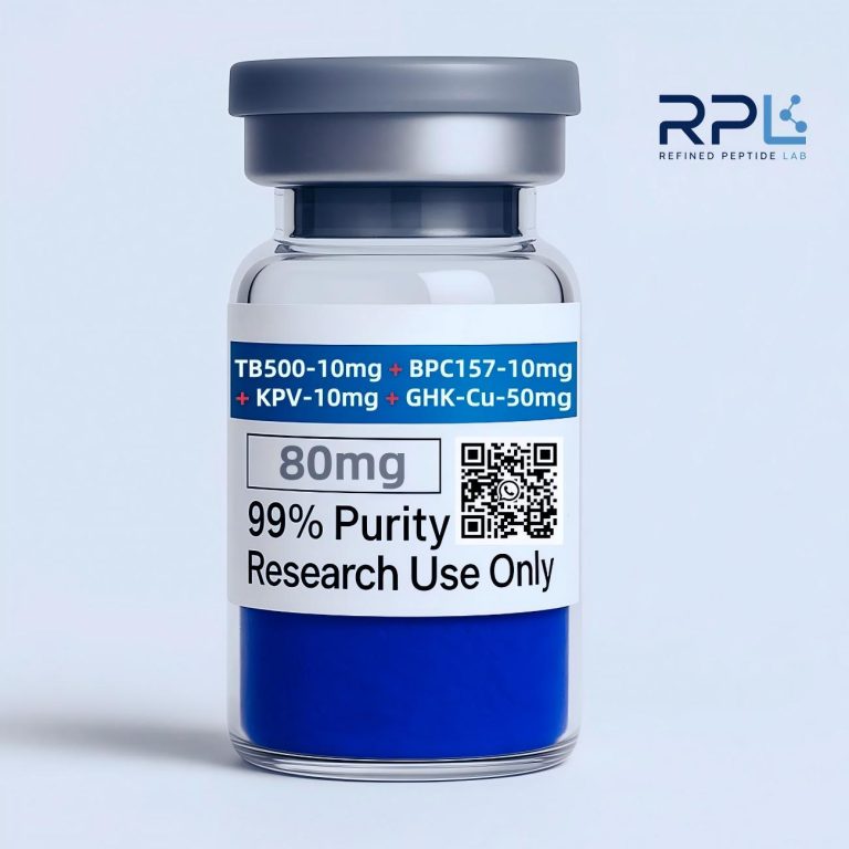
- KLOW80 Peptide Blend — Defined-stoichiometry KLOW blend, co-lyophilized for laboratory consistency.
15. Related Research Guides
- GHK-Cu Deep Dive — SPR binding kinetics, ECM remodeling, and SOD-mimetic activity of the copper tripeptide.
- TB-500 Deep Dive — Actin-sequestration biochemistry, G-actin binding, and cell migration data. BPC-157 + TB-500 is a common research pairing.
- Peptide Stability & Preservation Guide — Protocols for maintaining peptide integrity across conditions.
- How to Reconstitute Peptides — Step-by-step aseptic reconstitution and solubility optimization.
- Custom Peptide Synthesis & OEM — SPPS manufacturing, Fmoc chemistry, and bulk procurement standards.
Disclaimer: This article is intended exclusively for informational and research-context purposes. It does not constitute medical advice, product endorsement, or usage recommendation. BPC-157 is a research chemical that has not been evaluated by the FDA for safety or efficacy. All statements regarding biochemical mechanisms are based on published peer-reviewed literature and should be interpreted as research-context information, not as claims about any specific product’s effects. The significant concentration of BPC-157 research within a single laboratory group (§9) should inform the interpretation of all findings discussed herein. Individuals should consult appropriate regulatory and institutional guidelines before acquiring or using any research compound.